Oncological diseases are a problem of the 21st century. In almost all human organs and tissues, neoplasms can occur. After a thorough examination, specialists determine ways to get rid of them, identifying the degree of risk and type of tumor. A number of benign formations lend themselves to medical treatment, which cannot be said about malignant tumors. That is why the diagnosis of the disease plays an important role and determines the further viability of the organism as a whole and of a specific organ in particular. Learn how to diagnose pancreatic cancer.
How to identify the disease?
Most often, the pathology is determined by ultrasound. Pancreatic cancer may not manifest itself for a long time. Development occurs either against the background of a decrease in immunity, or as a result of complications of existing chronic organ diseases. Problem conditions in which a diagnosis of pancreatic cancer is necessary include diabetes mellitus and pancreatitis. In this case, experts recommend an annual ultrasound examination of the abdominal cavity, and if any factors that increase the risks are identified, donate blood to tumor markers.
Risk factors
In addition to the problems described above, directly related to pancreatic disorders, there are diseases that also increase the likelihood of a malignant tumor. These include:
- Smoking, giving up noticeably reduces the risk.
- Obesity, accompanied by an imbalance of sex hormones, can also be called a reversible factor. With a decrease in weight, adipose tissue leaves, which positively affects both the general condition and individual organs.
- Cirrhosis of the liver several times increases the possibility of an adverse outcome of any health problems.
- Allergic diseases of the skin, overgrown in a chronic form.
- Improper nutrition, which contains a large number of sausages, coffee, saturated fats, simple carbohydrates.
- Dental diseases.
Factors for which it is necessary to periodically diagnose pancreatic cancer include:
- Age over 60 years.
- The presence of oncopathology in the immediate family.
- Male affiliation.
- DNA mutations.
Symptoms
Pancreatic cancer manifestations are similar to some other diseases. Therefore, the average person may not attach importance to them for a long time. You should pay special attention to your health if you have the following conditions:
- Pain in the abdomen, in the hypochondrium and in the center, radiating to the back. At night and when leaning forward, it appears sharper, and subsides when the patient presses his legs to his stomach.
- Arising blood clots in the veins, visible to the naked eye.
- Jaundice, which is manifested first by yellowing of the skin, and then the integuments acquire a brown color with a green tint.
- The skin constantly itches due to stagnation of bile.
- Loss of appetite and weight loss.
- General weakness.
- Nausea and vomiting.
- Diarrhea, discoloration and odor of stool.
- Thirst, dry mouth.
- A large amount of urine with increased nighttime discharge.
- Discoloration of the mucous membranes and tongue.
- Dermatitis in the form of ulcers that pass by themselves and reappear, but in a different place.
- Swelling.
- Decreased libido.
- Signs of enlarged spleen, manifested by heaviness on the left in the hypochondrium.
- Hot flashes accompanied by heat on the face and body.
- Leg cramps.
Where to begin?
So, if you find yourself in a number of signs that indicate the occurrence of serious problems with the pancreas, it is imperative to visit a doctor. The specialist will begin the examination with a visual examination, collection of anamnesis and appoint to take tests. The early diagnosis of pancreatic cancer includes various laboratory tests that will help to understand whether there are problems with this organ or if the functions of others are impaired.
The tests prescribed for suspected cancer include:
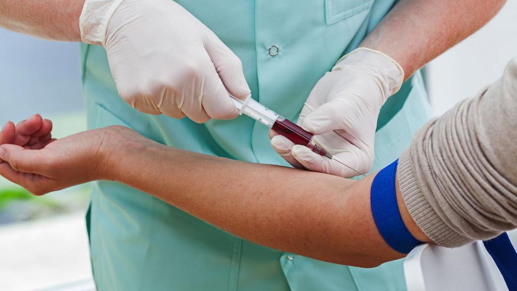
- Blood donation to CA-242 is carried out on an empty stomach with the exception of the use of sweet drinks on the previous day, all liquid is replaced with plain water. This is the main marker, which is a complex of protein and carbohydrate and secreted by the cells of the digestive system. A feature of the substance is its constant value in benign tumors and a significant increase in cancer pathology. If the figure approaches zero, then there are no pathologies, if it does not reach 20 units / ml, then you should know that this is how organ inflammation manifests itself. When the value is slightly higher, then additional studies are prescribed. An indicator that is too much more than 20 units / ml can indicate a malignant neoplasm in the stomach or pancreas. Tests for cancer, or rather, its suspicion detected in this way, include, in addition to CA-242, taking material on CA-19-9.
- Analysis for antigen CA-19-9 is prescribed precisely with the localization of the stomach and pancreas problems. CA-19-9 is a special substance released in cancer pathologies in an increased amount. However, experts argue that the data from this examination are not enough to make a diagnosis. If the analysis is repeated, since the cancer was detected earlier, and its value does not exceed 1000 units / ml, then they talk about the possibility of resection, that is, removal of part of the organ with the tumor. When the figure is more than 1000 units / ml, this in most cases means metastasis and the inability to cure.
- Diagnosing pancreatic cancer by blood involves determining the amount of pancreatic amylase. The so-called enzyme enters the pancreatic juice, which the pancreas produces, and travels to the intestine, where it breaks down carbohydrates. Most often, urine amylase analysis is added to this study. The norm of the first indicator should not exceed 53 units / ml, and the second - 200 units / ml. If cancer is suspected, the numbers can increase tens of times.
- Alkaline blood phosphatase is also required if laboratory diagnosis of pancreatic cancer is performed. This enzyme is involved in phosphorus-calcium metabolism, being an accelerator of chemical reactions. The norm in the blood is from 20 to 120 units / liter. The exception is newborns, pregnant women and patients over 75 years of age, whose rate is several times higher. In other cases, when determining a high value, they indicate the presence of a disease associated with stagnation of bile, including stage 4 pancreatic cancer.
- The delivery of feces for pancreatic elastase helps to distinguish a number of pathologies and differentiate the disease from other possible problems, such as cystic fibrosis and malabsorption. The norm is from 200 to 500 mcg / g.
To supplement the picture, experts and standard analyzes do not exclude it. In case of pancreatic cancer or suspicion of this disease, the doctor will definitely prescribe a laboratory study of both general blood counts and individual, such as the level of insulin, gastrin, glucagon, C-peptide.
Operation: Pros and Cons
Despite the fact that the differential diagnosis of pancreatic cancer is diverse and allows you to identify a lot of pathologies by submitting material for laboratory research, surgical intervention does not always confirm the development of a fatal disease.
The rationale for invading the body is the data obtained through clinical, instrumental and other types of analyzes. However, all of them can only to some extent indicate cancer. It is often impossible to determine an accurate diagnosis and distinguish chronic pancreatitis from an early stage of oncology, since benign tumors can exhibit similar symptoms and look identical. Only by the results of resection and research of the removed parts, it is possible with 100% probability to talk about pancreatic cancer. The 4th stage is the only stage that is unambiguously determined by radiation research methods, since it manifests itself by metastasis to the following organs:
- kidneys
- liver;
- lungs;
- intestines;
- spleen
- brain;
- bones.
Thus, the decision to conduct an operation is sometimes the only way to save a person’s life. Of course, the doctor pays special attention to the results of the tests and only in case of emergency it offers a resection. However, at the first stages of the examination, the role of tumor markers cannot be underestimated, according to which they determine the need for a thorough study and subsequent radiation diagnosis.
Instrumental methods
How to determine pancreatic cancer, or rather make sure the need for resection or to build a different treatment tactics, experts know. Preoperative methods for detecting pathology include:
- Ultrasound
- CT
- MRI
- ERCP.
- Hhhg
- PAT.
- Laparoscopy
- Biopsy.
Ultrasound procedure
When the manifestation of pancreatic cancer, symptoms that clearly indicate problems of this organ, begin to disturb the patient, he goes to the doctor. In the first stages of the examination of the patient, the specialist includes not only a survey and delivery of general tests, but also ultrasound of the abdominal cavity. Sometimes painful sensations indicate one organ, but in fact, another, located nearby. This method allows you to localize a possible focus of the disease and help the doctor to choose further methods of diagnosis or therapy.
An ultrasound scan may show an increase in any part of the pancreas or a change in its contour. Particular attention in ultrasound is given to the head of the gland, since in 80% of cases it is in it that a new formation is observed. In the caudal part, cancer is much less likely to manifest itself. However, it happens that the examination reveals a tumor of the entire tissue, which in fact may not be an oncological disease, but an acute form of pancreatitis.
Ultrasound also helps to visualize the nature of the changes and the structure of the gland. Usually, with this form of cancer, the tumor is hypoechoic and does not have internal echostructures.
CT scan
This study is conducted using x-rays that pass through organs and tissues. Since they all have different densities, as well as cancer formation, the apparatus manages to transmit the image in layers. The final display allows you to visualize those organs that have undergone tomography, and their structure. The specialist can evaluate not only the size of the pancreas, but also various deposits, inflammations and swelling. It should be noted that the degree of emission of QDs is much less than that of ordinary X-rays. When preparing for this type of examination, you need to keep in mind that contrasting is often used. Therefore, the presence of contraindications to the use of iodine-containing preparations must be voiced by the attending physician. You should also notify your doctor of any allergic reactions you have to medicines.
Magnetic resonance imaging
This is a well-established method based on magnetic radiation. It gives complete information about tissues, since it is carried out by exposing the body to a magnetic field. As a result, the vibration of atoms in human cells allows a special program to create a three-dimensional image, which is much better than two-dimensional images. The examination is carried out in the supine position, when the patient is motionless, and magnetic coils and the detector of the apparatus rotate around him. In a few minutes, about a hundred photographs are taken in different planes, providing an image through software processing, and a radiologist describes the state of the organ under examination and hands out a disk with the results of magnetic resonance imaging of the pancreas.
Endoscopic retrograde cholangiopancreatography
The method works using a contrast medium. It can be called combined, because it combines endoscopic and radiological examination. An endoscope is inserted into the duodenum. Through it, a special preparation is served in the papilla papilla, and then several pictures are taken.
The use of high-tech equipment allows tracking of the process at all its stages, and the method also has a small exposure. The quality of the cholangiopancreatogram allows us to judge the problems of the pancreas and bile ducts with a high degree of accuracy.
Percutaneous transhepatic cholangiography
This method also represents a fluoroscopic examination using an iodine-containing substance. Unlike the previous version, the drug enters through the skin. The patient is placed on an x-ray table and fixed.
The place where they plan to insert the needle is treated and separated from the rest of the surface with sterile materials, after which a local anesthetic is injected. As the patient exhales, they are asked to hold their breath and insert a needle into the intercostal space. Having penetrated the liver parenchyma, the needle is slowly removed, while releasing a contrast medium until the bile duct is detected, into which the remaining preparation is injected. The equipment screen allows you to evaluate the filling of the ducts, after which they take several pictures.
Positron Emission Tomography
In this case, a substance that performs the function of contrast is injected into a vein. The difference from previous methods is the use of sugar labeled with isotope. Here, the study is based on the ability of cancer cells to accumulate radioactive substances. In the images, malignant tumors, if any, will significantly differ in color from the rest of the tissues, which will allow them to be localized and decide on further therapy or surgery.
Laparoscopy
As a surgical method, it is prescribed if necessary to exclude the presence of cancer cells in the pancreas. A resection of a benign tumor is also performed. Removal of malignant tumors in this way is unacceptable.
During this procedure, despite tiny incisions, the patient needs anesthesia. In this case, anesthesia-air composition is chosen, which is fed through a special tube. The essence of the technique of surgical intervention is the implementation of three to four small incisions, after which carbon dioxide is pumped into the abdominal cavity. Then, through the punctures, the tools are introduced and the necessary manipulations are performed.
If the examination reveals that laparoscopy will not help, surgeons may decide to have an open laparotomy.
Biopsy
The most difficult for the patient and at the same time the most informative method is a biopsy of pancreatic cancer. Such a study involves excising a portion of the tissue or taking a small number of cells for subsequent examination with a laboratory microscope. After taking the tissue, it is stained with a special composition and histological examination is performed.
There are 4 ways to take cells:
- Intraoperative, when cells are obtained through ordinary laparotomy. A direct, transduodenal and aspiration fine-angle biopsy can be used here.
- Laparoscopic, the material is taken at which, making small incisions.
- Percutaneous, where cells for research are obtained under the supervision of ultrasound and CT. It is this method from all of the above that is considered the safest and less traumatic, but it can not always be used.
- An aspiration biopsy is used in most cases where material can be taken. The accuracy of the study is 96%.
Patients who have been diagnosed with oncology based on the results of analyzes and studies should be aware that this is not a sentence.
Firstly, it happens that after resection and subsequent histology, it is revealed that the result was false positive. And this means that the excised tissue might not be an oncological formation, but was a benign tumor.
Secondly, the decision on surgical intervention is made by a specialist. Therefore, after examinations, find a good doctor and read reviews about him.
Thirdly, after getting rid of malignant tissues, you can live happily ever after.
Remember that diagnosing cancer at an early stage is half the success. Watch your health and consult a doctor if you see any bad symptoms.