In the anatomy of both humans and animals, the notion of “brachiocephalic trunk” stands out. Today we’ll talk about this in more detail.
General concept
The name of this body speaks for itself. The brachiocephalic trunk moves from the aorta along the midline of the sternum. Then it rises up obliquely, then back and up, and at the level of the clavicular joint it is divided into two arteries. It is located in front of the trachea, which in children is covered with a thymus gland, and has a short length of three to four centimeters.
In infants, he often divides cephalad into sternoclavicular articulation in the anterior triangle of the neck.
Human trunk
In humans, this organ has the structure described above. This, as a rule, is a short and thick vessel that branches into two right arteries, which are covered by pleura on both sides - to the right and front. In the left part of the human body , this artery is not. Otherwise, this vessel is called the brachycephalic stem (from the Latin name) or the nameless artery.
In a person, some diseases may be associated with a brachiocephalic trunk. Among them are the following:
- atherosclerosis (accumulation of cholesterol and fat on the inner walls of arteries);
- congenital malformations;
- hemangiomas (a benign tumor that develops from small blood vessels);
- arterial injury;
- aneurysms (lumen widening two or more times);
- obliterating lesions of the branches of the arc (impaired vascular patency, which leads to ischemia of the brain and limbs (upper)).
If there are problems with this vessel, you should contact an angiosurgeon.
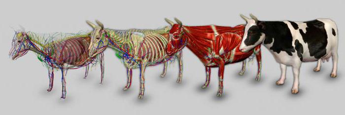
Trunk in animals
The anatomy of the brachiocephalic trunk is as follows. It differs from the human in that it goes to the entrance to the chest cavity and already there it divides into two left arteries (in humans, the right ones). This occurs at the level of the second thoracic vertebra.
Some animals, for example, a dog and a pig, do not have a brachiocephalic trunk; instead, they have two left arteries that exit from the aortic arch. From one of the arteries, which is called the brachiocephalic, carotid arteries depart, which carry blood to the head of animals. An exception is a horse, in which several more small arteries depart from it.
The brachiocephalic trunk provides blood to the head, neck, chest limbs, and part of the chest wall.
In some cases, the lower thyroid artery goes from this trunk to the lower part of the thyroid gland. Thanks to the trunk, the deficiency or absence of one of the vessels of the thyroid gland can be compensated.
Trunk branches
In the order of departures of the vessels from the subclavian arteries, there are species-specific characteristic differences. The following branches extend from the arteries:
- The rib-cervical trunk supplies blood to the muscles of the neck and withers. It departs together with such arteries as the deep cervical and vertebral (in ruminants and pigs), or only the first of them (in carnivores). In horses, this trunk is an independent branch.
- The deep cervical artery supplies the extensors of the head and neck. Diverges in the cervical muscles, its direction is cranial. On the neck, like the vertebral branch, forms the 2nd collateral. In pigs and dogs, this artery is a branch of the costal cervical trunk.
- The vertebral artery is paired. It is also directed cranially. When he reaches the atlas, he releases the branches into the muscles and the spinal cord, exits through the hole in the first cervical vertebra (atlas) of the animals and forms large blood flow paths (called collateral) on the neck. In cattle leaves with the above branches. And in carnivores, it is the first blood vessel that departs from the subclavian artery.
- The bald artery (also called the superficial cervical artery) provides blood to the muscles of the neck, chest, and also the entrance to the chest. In a pig, the thyroid trunk departs from it.
- Internal and external thoracic arteries. The inner one is directed caudally along the surface of the sternum, reaches the seventh rib and branches. Its final vessel is represented by the muscular-diaphragmatic artery. Then it goes down and supplies blood to the muscles of the abdominal cavity, in pigs and carnivores also the mammary gland. The external artery bypasses the first rib and branches deep in the pectoral muscle. This artery is quite poorly developed.
Further, the brachiocephalic trunk of the animals, continuing the left arteries, becomes axillary arteries. They are the main source of blood supply to the chest limbs.