Today we will talk about a rather rare childhood disease, accompanied by a large number of abnormalities and developmental disorders. It will be about Edwards syndrome. We will analyze its causes, forms, frequency of manifestation, diagnostic methods and other important issues.
What is it?
Edwards syndrome is a disease caused by chromosomal abnormalities, causing a list of disorders and abnormalities in the development of the child. Its cause is trisomy of the 18th chromosome, i.e., the presence of its extra copy. This fact leads to the appearance of complications of a genetic nature.
The risk that a child with Edwards syndrome is born is 1 out of 7,000 cases. Unfortunately, most babies with this deviation die in the first weeks of life. Only about 10% live one year. The disease leads to deep mental retardation, congenital lesions of internal and external organs. The most common of these are defects in the brain, heart, kidneys, small head and jaw, cleft lip or palate, clubfoot.
The symptoms of the disease were first formed and described in 1960 by D. Edwards. The doctor managed to establish a relationship between the manifestation of several signs, found more than 130 defects that accompany the disease. Although the symptoms of Edwards syndrome are very pronounced, modern methods of therapy against them, unfortunately, are powerless.
Cause of disease
If Edwards syndrome (photos of sick children for ethical reasons will not be posted) was diagnosed during pregnancy, then most often the latter ends in a miscarriage or stillbirth. Alas, the manifestation of the disease in the fetus cannot be prevented today.
Also, in the modern world, the clear causes of this genetic disease have not been elucidated, and therefore preventive measures against its development in future children cannot be formed. However, experts identified risk factors:
- Adverse environmental conditions.
- The effects of radiation, toxic, chemicals on parents.
- Addiction to alcohol and tobacco.
- Heredity.
- Taking certain medications.
- Incest, consanguinity of parents.
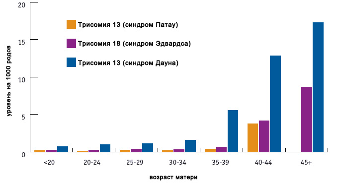
- The age of the future mother. If a woman is older than 35 years old, then this is considered the cause of Edwards syndrome in the child, as well as other chromosomal diseases.
Syndrome Forms
The type of such anomaly is primarily affected by the stage of development of the embryo, on which the embryo overtakes the syndrome. There are three types of differences:
- Full. The most severe type, it accounts for 80% of cases. A triple chromosome appears at a time when the fetus was just one cell. It follows that the abnormal chromosome set will be transmitted during division and to all other cells, observed in each of them.
- Mosaic. The name is given due to the fact that healthy and mutated cells are mixed like a mosaic. 10% of those affected by Edwards's symptom suffer from this particular form. Signs of the disease are less pronounced here, but still interfere with the normal development of the child. An excess chromosome appears during the phase when the embryo consists of several cells, so only a part of the body or an individual organ is affected.
- Possible translocation. Here, not only chromosome nondisjunction is observed, but also an overabundance of information generated by translocation restructuring. It manifests itself both during the maturation of gametes and during the development of the embryo. Deviations are not pronounced here.
Syndrome prevalence
The risk of Edwards syndrome cannot be expressed in exact numbers. The lower boundary of the birth of a child with such an anomaly is 1: 10000, the upper one is 1: 3300. At the same time, it occurs 10 times less than Down syndrome. The average fertility rate for children with Edwards disease is 1: 3000.
According to studies, the risk of giving birth to a baby with this syndrome increases with a parent's age of more than 45 years by 0.7%. But it is also present in 20-, 25-, 30-year-old parents. The average age of the father of a child with Edwards syndrome is 35 years, and that of a mother is 32.5 years.
The anomaly is also related to gender. It is proved that in girls it occurs 3 times more often than in boys.
Syndrome and pregnancy
Edwards syndrome is still showing signs of pregnancy. The latter proceeds with a number of complications, it is characterized by overshoot - babies are born at about 42 weeks.
At the stage of pregnancy, the fetal disease is characterized by the following:
- Inadequate activity of the embryo.
- Bradycardia - a reduced heart rate.
- Polyhydramnios.
- Discrepancy between the size of the placenta and the size of the fetus - it has a smaller size.
- The development of one umbilical artery instead of two, which leads to oxygen deficiency, asphyxiation.
- Abdominal hernia.
- Plexus of vascular formations, visible on ultrasound (found in 30% of children affected by the syndrome).
- Underweight fetus.
- Hypotrophy is a chronic disorder of the gastrointestinal tract.
60% of children die in the womb.
Antenatal diagnosis
Ultrasound Edwards syndrome can only be determined by indirect signs. The most accurate method for diagnosing a fetal syndrome today is perinatal screening. Based on it, in case of alarming suspicions, the doctor already sends the woman for invasive testing.
Screening revealing a karyotype of Edwards syndrome is divided into two stages:
- The first is carried out at 11-13 weeks of gestation. Biochemical parameters are studied - the mother's blood is checked for hormone levels. The results at this stage are not final - they can only talk about the presence of risk. For calculations, a specialist needs protein A, hCG, a protein produced by the membranes of the embryo and placenta.
- The second stage is already aimed at an accurate result. For research, a cord blood or amniotic fluid sample is taken, which is then subjected to genetic analysis.
Invasive testing
Chromosomes of Edwards syndrome are most likely to be determined by this method. However, it necessarily involves surgical intervention and penetration into the shell of the embryo. Hence the risk of abortion and the development of complications, which is why the test is prescribed only in extreme cases.
Three types of sampling are known today:
- BVH (chorionic villus sampling). The main advantage of the method is that a sample is taken starting from the 8th week of pregnancy, which allows one to identify complications in the early stages. For research, you need a chorion sample (one of the layers of the placental membrane), the structure of which is similar to the structure of the embryo. This material allows you to diagnose intrauterine infections, genetic and chromosomal diseases.
- Amniocentesis. The analysis is carried out starting from the 14th week of pregnancy. In this case, the amniotic membrane of the embryo is pierced with a probe, the instrument collects a sample of amniotic fluid containing the cells of the unborn child. The risk of developing complications from such a study is much higher than in the previous case.
- Cordocentesis. The term is not earlier than the 20th week. Here, a fetal cord blood sample is taken. The difficulty is that when taking the material, the specialist has no right to make a mistake - he must get the needle exactly into the umbilical cord vessel. In practice, this happens as follows: a puncture needle is inserted through the front wall of the peritoneum of a woman, which collects about 5 ml of blood. The procedure is controlled by ultrasound devices.
All of the above methods can not be called painless and safe. Therefore, they are carried out only in cases where the risk of a genetic disease in the fetus is higher than the risk of developing complications from taking material for analysis.
Parents need to remember that the mistake of a physician during the procedure can lead to the manifestation of serious diseases, congenital malformations in the unborn child. The risk of abortion from such an intervention cannot be ruled out.
Non-invasive testing
Diagnosis of Edwards syndrome in the fetus includes non-invasive methods. That is, without penetration into the membranes. Moreover, such methods are not inferior to invasive ones.
One of the high-precision analyzes of this type can be called karyotyping. This is a sample of the mother’s blood, which contains the free DNA of the embryo. Experts extract them from the material, copy, and then carry out the necessary studies.
Postpartum diagnosis
A specialist can identify children with Edwards syndrome and externally. However, for the final diagnosis, the following procedures are performed:
- Ultrasound - a study of pathologies of internal organs, necessarily the heart.
- Tomography of the brain.
- Consultation of a children's surgeon.
- Examination by specialists - an endocrinologist, neurologist, otolaryngologist, ophthalmologist, who previously worked with children suffering from this disease.
Abnormalities in the syndrome
Pathologies, the cause of which is trisomy on the 18th chromosome, are quite serious. Therefore, only 10% of children with Edwards syndrome survive up to a year. Basically, girls live no more than 280 days, boys - no more than 60.
In children, the following external deviations are observed:
- An elongated skull.
- Microcephaly (small head and brain size).
- Hydrocephalus (fluid buildup in the cranium).
- Narrow forehead with wide nape.
- Abnormally low ears. There may be no lobe or tragus.
- Shortened upper lip, making the mouth triangular.
- High sky, often with a gap.
- Deformed jaw bones - the lower jaw is abnormally small, narrow and undeveloped.
- Cropped neck.
- Abnormally narrow and short palpebral fissures.
- The absence of a part of the eye membrane, cataract, coloboma.
- Impaired joint function.
- Underdeveloped, inactive feet.
- Due to the abnormal structure of the fingers, fins-like limbs may form.
- Heart disease.
- Abnormally enlarged chest.
- Disturbed work of the endocrine system, in particular, the adrenal glands and the thyroid gland.
- Unusual bowel location.
- Irregular kidney shape.
- Doubling of the ureter.
- Boys have cryptorchidism, girls have a hypertrophied clitoris.
Mental abnormalities are usually as follows:
- Underdeveloped brain.
- Complicated degree of oligophrenia.
- Convulsive syndrome.
The prognosis for patients with Edwards syndrome
Unfortunately, the forecasts today are disappointing - about 95% of children with this disease do not live up to 12 months. Moreover, the severity of its form does not depend on the ratio of sick and healthy cells. Surviving children have physical abnormalities and a severe degree of oligophrenia. The vital activity of such a child needs comprehensive monitoring and support.
Often children with Edwards syndrome (photos are not presented in the article for ethical reasons) begin to distinguish the emotions of others, smile. But their communication, mental development is limited. Over time, a child can learn to raise his head, eat.
Therapy options
Today, such a genetic disease is incurable. The child is prescribed only therapy that supports his condition. The patient’s life is associated with many anomalies and complications:
- Muscle atrophy.
- Strabismus.
- Scoliosis.
- Malfunctions of the cardiovascular system.
- Atony of the intestine.
- Low tone of the walls of the peritoneum.
- Otitis.
- Pneumonia.
- Conjunctivitis.
- Sinusitis.
- Diseases of the genitourinary system.
- High probability of developing kidney cancer.
Conclusion
Summing up, I want to note that Edwards syndrome is not inherited. Patients in most cases do not live to reproductive age. However, they are not capable of procreation - the disease is characterized by underdevelopment of the reproductive system. As for the parents of a child with Edwards syndrome, the chance of making the same diagnosis during the next pregnancy is 0.01%. I must say that the disease itself manifests itself very rarely - it is diagnosed only in 1% of newborns. There are no special reasons for its occurrence - in most cases, parents are completely healthy.