The vas deferens is a paired organ that is part of the vas deferens of the appendage and testicles, as well as an integral part of the epididymis. This duct ends at the junction with the canal of the seminal vesicle.
The vas deferens is one of the main management tools for the male reproductive system. The final part of it forms an ampoule in the form of a spindle, which is part of the prostate gland and converges with the excretory channel of the seminal vesicle. The unifying duct is called the vas deferens.
Length
The length of the vas deferens is 45 - 50 centimeters. In the transverse section, it does not exceed three millimeters, and the diameter of the lumen is not more than half a millimeter. The walls of the duct are significantly thickened, and in this regard, it is easily palpated on the surface of the spermatic cord from the scrotum to the ring of the inguinal canal.
The anatomy of the vas deferens is of interest to many, so let's take a closer look at its structure.
Four duct sections
Based on the topographic data of the vas deferens, four of its departments are distinguished:
- The first section is called the initial (shortened testicle). It is located behind the testicle, closer to its appendages. This is the smallest section located at the back of the testicle.
- Further, if you climb cranially (vertically), it is followed by the funicular section. It is located within the spermatic cord, closer to the middle part from its vessels, and stretches to the inguinal ring located on the surface. It should be noted that the structure of the vas deferens is unique.
- After this, the duct enters the inguinal canal (inguinal part). Leaving from which, it stretches through the inguinal ring, goes through the small pelvis, and more specifically, through its side wall to the lower part until it connects to the excretory channel of the seminal vesicle. This section of the duct is called the pelvic. The pelvic section (pars pelvina) starts from the inside of the opening of the inguinal canal and ends with the prostate gland. It is devoid of vascular plexus and extends through the parietal sheet of the peritoneal part of the pelvis. The final part of the duct carrying the seed is located near the bottom of the bladder and becomes wider, resembling an ampoule.
- The duct of the vas deferens in the pelvic area is located extraperitoneally in the retroperitoneal space (that is, only in one part). Towards the prostate gland from the lateral side (side), it bypasses the rod of the lower epigastric artery, connects to the iliac artery and vein, passes between the rectum and the bladder, crosses with the ureter, gets to the bladder and reaches the base of the prostate gland, being near such same duct on the other hand. This final part of the vas deferens is enlarged, resembles a fusiform shape and forms an ampoule of the vas deferens.
The length of the ampoule is 30-40 millimeters, and its largest transverse dimension reaches ten millimeters. In the lower distal (most distant) part of the vessel, it gradually becomes narrow, penetrating into the thick layer of the prostate gland and connecting with the excretory duct of the seminal vesicle.
The single duct is called the ejaculatory. Two of them enter the prostate area of the urethra near the seminal tubercle and extend to the lower part through the posterior region of the prostate. The length of each of the ejaculatory ducts is 2 cm. The internal diameter is 1 mm in its original part and 0.3 mm at the point of entry into the urethra.
Wall structure
The wall of the duct that carries the seed is formed by the mucosa, muscle and adventitia membranes. The first of them is three to five longitudinal folds. At the site of the vessel of the described duct, the mucous membrane forms coiled tubercles, which are called ampoule diverticula.
The muscular membrane is located in the outer part of the mucosa, it is formed through the inner, middle circular and outer longitudinal layers of smooth muscle cells. The muscular membrane supplies the wall of the vas deferens with almost cartilaginous density. The muscle membranes of the vessel of this duct are not so clearly represented. On the outside, its wall is formed by the adventitia membrane, which smoothly passes into the connecting layer of the surrounding duct.
Duct designation
In the vas deferens duct, mature, immobile sperm with a fluid having an acidic environment, as a result of the reduction of the duct wall, leave the epididymis and are stored in the duct vessel. It should be noted that the liquid there is partially absorbed.
The provision of the duct and seminal vesicle with nerve cells is sympathetic (this system is formed from the upper and lower hypogastric plexuses), as well as parasympathetic (through the pelvic internal nerves).
Duct blood supply
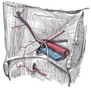
Blood supply to the vas deferens (photo presented in the article) is due to the ascending branch of the artery, the middle rectal artery and lower urinary bladder.
Blood supply to the seminal vesicle is also formed by the branches of the upper and middle rectal arteries, as well as the lower urinary bladder artery.
The veins of the seminal vesicles of the male reproductive system enter the plexus of the veins of the bladder, and the veins of the vas deferens flow into the tributaries of the internal iliac vein.
Physiology of seminal vesicles
Seminal vesicles are glandular androgen-dependent organs, the secretion of which consists of a viscous, white-gray jelly-like substance, which after ejaculation becomes liquid in a few minutes and forms 50-60 percent of sperm. The main function of the seminal vesicles is manifested in the secretion of fructose, the level of which reflects the androgenic saturation of the body.
Seminal vesicles secrete other components of sperm, namely:
- nitrogenous substances;
- inositol;
- proteins;
- ascorbic acid;
- prostaglandins.
The excretion of seminal vesicles together with the secretion of the testicles is a protective colloid, creating greater resistance for sperm.