What is the principle of the x-ray laser? Due to the high gain in the lasing medium, the short lifetimes of the upper state (1-100 ps) and the problems associated with the construction of mirrors that can reflect the rays, these lasers usually work without mirrors. An X-ray beam is generated in one pass through the amplification medium. The emitted radiation based on the amplified spontaneous beam has a relatively low spatial coherence. Read the article to the end and you will understand that this is an X-ray laser. This device is very practical and unique in its structure.
Kernels in the structure of the mechanism
Since conventional laser transitions between visible and electronic or vibrational states correspond to energies up to 10 eV, various active media are needed for X-ray lasers. Again, various active charged nuclei can be used for this.
Weapon
Between 1978 and 1988, in the Excalibur project, the US military attempted to develop an X-ray laser with a nuclear explosive defense device for missile defense as part of the Star Wars Strategic Defense Initiative (SDI). The project, however, turned out to be too expensive, dragged on and was eventually frozen.
Plasma media inside the laser
The most commonly used media include highly ionized plasma created in a capillary discharge, or when a linearly focused optical pulse hits a solid target. According to the Saha ionization equation, the most stable electronic configurations are neon, with the remaining 10 electrons, and nickel-like with 28 electrons. Electron transitions in a highly ionized plasma usually correspond to energies of the order of hundreds of electron-volts (eV).
An alternative amplifying medium is the relativistic electron beam of a free electron x-ray laser, which uses stimulated Compton scattering instead of standard radiation.
Application
Applications for coherent X-ray radiation include coherent diffraction imaging, the study of dense plasma (opaque to visible radiation), X-ray microscopy, phase-resolution medical imaging, material surface research, and use as weapons.
A lightweight version of the laser can be used for ablative laser movement.
X-ray laser: how it works
How do lasers work? Due to the fact that a photon enters an atom with a certain energy, you can make an atom emit a photon with this energy in a process called stimulated radiation. By repeating this process on a large scale, you get a chain reaction that leads to a laser. However, some quantum sites make this process stop, because the photon is sometimes absorbed without radiation at all. But to provide maximum chances, photon energy levels are increased, and mirrors are placed parallel to the light path to help scattered photons return to the game. And at high X-ray energies, special physical laws are found that are characteristic of this particular phenomenon.
History
In the early 1970s, the X-ray laser seemed out of reach, since most of the lasers of that time reached a peak at 110 nm, which is significantly less than that of the largest X-rays. This was due to the fact that the amount of energy needed to obtain the stimulated material was so high that it had to be delivered with a fast pulse, which further complicated the reflection ability needed to create a powerful laser. Therefore, scientists looked at the plasma, because it looked like a good conducting medium. A team of scientists in 1972 stated that they had finally achieved the use of plasma in the development of lasers, but when they tried to reproduce the previously obtained results, for some reason nothing came of it.
In the 1980s, a major player from the world of science, Livermore, joined the research team. Scientists, meanwhile, have taken small but important steps over the years, but after the Defense Advanced Research Projects Agency (DARPA) stopped paying for X-ray research, Livermore became the leader of the research group. He led the development of several types of lasers, including those created on the basis of synthesis. Their nuclear weapons program was promising, because the high-energy indicators that scientists achieved during this program hinted at the possibility of creating a high-quality pulsed mechanism that would be useful in the construction of an X-ray free electron laser.
The project was gradually approaching completion. Scientists George Chaplin and Lowell Wood first studied fusion technology for X-ray lasers in the 1970s, and then switched to the nuclear version. Together they developed such a mechanism and were ready for testing on September 13, 1978, but a hardware failure interrupted it. But perhaps it was for the better. Peter Hagelstein created a different approach after studying the previous mechanism, and on November 14, 1980, two experiments proved that the prototype X-ray laser worked.
Star Wars Project
The US Department of Defense became interested in the project very soon. Yes, using the power of a nuclear weapon in a focused beam is too dangerous, but this force could be used to destroy intercontinental ballistic missiles (ICBMs) in the air. It would be most convenient to use a similar mechanism in near-Earth orbit. The whole world knows this program called Star Wars. Nevertheless, the project of using an X-ray laser as a weapon was not crowned with success.
The February 23, 1981 issue of Aviation Week and Space Technology magazine presented the results of the project’s first tests, including a laser beam whose pulse wave reached 1.4 nanometers and hit 50 different targets.
The tests of March 26, 1983 yielded nothing due to a sensor failure. However, the following tests of December 16, 1983 demonstrated its true capabilities.
The fate of the project
Hagelstein suggested a two-stage process in which the laser created a plasma that would release charged photons that collide with the electrons of another material and cause the emission of x-rays. Several installations were tested, but in the end, ion manipulation was the surest solution. The plasma removed the electrons until there were only 10 internal electrons left, where the photons then charged them to the 3p state, thereby releasing the “soft” beam. The July 13, 1984 experiment proved that it was more than a theory when a spectrometer measured strong emissions at 20.6 and 20.9 nanometers of selenium (a neon-like ion). Then the first laboratory (non-military) X-ray laser with the name Novett appeared.
The Fate of Novett
This laser was developed by Jim Dunn and had physical aspects tested by Al Osterheld and Slava Shlyaptsev. Using a fast (near nanosecond) high-energy light pulse that charged particles to emit X-rays, Novett also used glass amplifiers that increase efficiency but also heat up quickly, which means that it can only work 6 times a day between cooling. But some work has shown that it can shoot a picosecond pulse, while compression returns to a nanosecond pulse. Otherwise, the glass amplifier will be destroyed. It is important to note that Novett and other “desktop” X-ray lasers create “soft” X-rays that have a longer wavelength, which prevents the beam from passing through many materials, but gives an idea of the alloys and plasma, since it is easy to see them through.
Other applications and functional features
So what can you use this laser for? It was previously noted that a shorter wavelength may facilitate the study of some materials, but this is not the only area of application. When a target is struck by an impulse, it is simply destroyed by atomic particles, and the temperature in this case reaches millions of degrees in just a trillionth of a second. And if this temperature is enough, the laser will cause the electrons to exfoliate from the inside. This is due to the fact that the lowest level of electronic orbitals implies the presence of at least two electrons that are emitted from the energy generated by x-rays.
The time taken for an atom to lose all its electrons is of the order of several femtoseconds. The resulting nucleus does not linger for a long time and quickly passes into the plasma state, known as "warm dense matter", which is mainly found in nuclear reactors and the nuclei of large planets. By conducting experiments with the laser, we can get an idea of both processes, which are various forms of nuclear fusion.
The use of an X-ray laser is truly universal. Another useful property of these X-rays is their use with synchrotrons or particles accelerating along the path of the accelerator. Based on how much energy is required for this path, particles can emit radiation. For example, electrons emit x-rays when excited, which have a wavelength near the size of an atom. Then we could study the properties of these atoms through interaction with x-rays. In addition, we can change the electron energy and obtain different wavelengths of x-rays, achieving greater depth of analysis.
However, it is very difficult to create an x-ray laser with your own hands. Its structure is extremely complex even from the point of view of experienced physicists.
In biology
Even biologists were able to benefit from nuclear-pumped x-ray lasers. Their radiation can help reveal aspects of photosynthesis previously unknown to science. They capture subtle changes in the leaves of plants. Long waves of soft x-rays of the laser allow you to explore without destruction everything that happens inside the plant. The nanocrystal injector triggers photocell I, the protein key to photosynthesis necessary for its activation. This is intercepted by a laser beam of x-rays, which causes the crystal to literally explode.
In case of further success of the above experiments, people will be able to unravel the secrets of nature, and artificial photosynthesis can become a reality. There will also be a question about the possibility of more efficient use of solar energy, provoking the appearance of scientific projects for many years to come.
Magnets
How about an electronic magnet? Scientists found that when they had xenon atoms and molecules limited by iodine, hit by a high-power x-ray, the atoms discarded their internal electrons, creating a void between the nucleus and the most distant electrons. The forces of attraction set these electrons in motion. Usually this should not happen, but due to the sudden drop of electrons, an overly “charged” situation at the atomic level occurs. Scientists think that laser can be used in image processing.
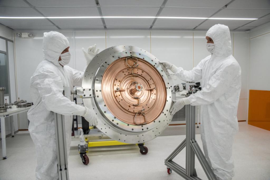
Giant Xfel Laser
Located at the US National Accelerator Laboratory, in particular a linear accelerator, this 3,500-foot laser uses several ingenious devices to hit targets with hard X-rays. Here are some of the components of one of the most powerful lasers (abbreviations and English indicate the components of the mechanism):
- Drive Laser - creates an ultraviolet pulse that removes electrons from the cathode. It emits electrons to an energy level of 12 billion eW by manipulating the electric field. Also inside the mechanism there is an S-shaped accelerator called Bunch Compressor 1.
- Bunch Compressor 2 is the same concept as in Bunch 1, but a longer S-shaped structure, enlarged due to higher energies.
- Transport Hall - allows you to make sure that the electrons are suitable for focusing pulses using magnetic fields.
- Undulator Hall - consists of magnets that make electrons move back and forth, thereby generating high-energy x-rays.
- Beam Dump is a magnet that picks up electrons but transmits x-rays without movement.
- LCLS Experimental Station is a special chamber in which the laser is mounted and which is the main space for experiments related to it. The rays generated by this device generate 120 pulses per second, with each pulse lasting 1/10000000000 of a second.
- Capillary plasma-discharge medium. In this setup, a few-centimeter-long capillary made of a stable material (such as alumina) limits a high-precision, sub-microsecond electrical pulse in a low-pressure gas. The Lorentz force causes further compression of the plasma discharge. In addition, a pre-ionization electric or optical pulse is often used. An example is a capillary neon-like Ar8 + laser (generating radiation at 47 nm).
- The target medium of a continuous plate is that after a hit by an optical pulse, the target emits a highly excited plasma. Again, a longer “pre-pulse” is often used to create the plasma, and a second, shorter and more energetic pulse is used to further heat the plasma. For short lifetimes, an impulse shift may be required. The gradient of the plasma refractive index causes the amplified pulse to bend from the target surface, since at frequencies above resonance the refractive index decreases with the density of the substance. This can be compensated for by using several targets in a series of emissions, as in the European free electron x-ray laser.
- Plasma excited by an optical field - at optical densities high enough for efficient tunneling of electrons or even to suppress a potential barrier (> 1016 W / cm2), gas can be strongly ionized without contact with a capillary or target. A collinear setting is usually used to synchronize pulses.
In general, the structure of this mechanism is similar to a European free electron x-ray laser.