The sacral plexus (Latin name - plexus sacralis) is formed by the 4th and 5th abdominal branches of the lumbar and spinal sacral nerves. They are formed into a bundle called the lumbosacral trunk (in Latin - truncus lumbosacralis) and is part of plexus sacralis. This plexus includes fibers from the nodes of the lower lumbar and sacral sympathetic trunk. Sacral plexus branches are located on the piriformis muscle (Latin name m. Piriformis) in the pelvis and converge to the holes located above and below the piriformis muscle. Through the above holes, the branches go to the back of the pelvis.
Plexus with short mixed branches
The spine for a person is functionally important. Due to the lumbar vertebra, the formation of lordosis occurs. This section of the spine is experiencing the greatest load.
The sacral plexus is located anterior to the transverse processes of the lumbar vertebrae. Its anatomy is unique and has been studied for a long time.
Muscle branches
The muscle branches (Latin name - rr. Musculares) are formed by fibers L 4 and L 5, as well as S 1 and S 2 , supply the nerves of the pelvic region m. piriformis, obturatorius internus. After passing through the hole under the piriformis muscle, they bind the quadriceps femoral muscle (m. Quadratus femoris) to the central nervous system. Receptors of other fibers are present in these soft tissues. For example, femoral nerve tissue.
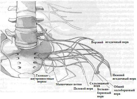
Upper buttock
The superior gluteal nerve (in Latin - n.gluteus superior) is formed by fibers L 2 - L 5 and S 1 and is represented by a short trunk. It follows through the supra-pear-shaped opening from the small pelvis to the back of the pelvis. It is combined into a joint bundle with arteries and a vein of the same name. The nerve is divided into 3 branches, which supply the small and medium muscles of the buttocks and thighs with sensitive fibers. Receptors are located in the small, medium muscle tissue and connective membrane. The nerves of the sacral plexus are important.
Lower buttock
The lower gluteal nerve (Latin name n.gluteus inferior), which is formed by the fibers L 5 and S 1 -S 2 , is represented by a short trunk extending to the back of the pelvis through the slit-like gap in the lower part of the large paired hole of the posterior lower pelvic wall, as and blood vessels. The large lumbar muscle is supplied with nerves. Receptors are located both in the hip joint and in the gluteus maximus muscle. There is a connection of the fibers of the sensory nerve and motor fibers. Then they move together to the nuclei of the spinal cord.
Sacral plexus and long branches
The muscle branches extend from all the anterior branches that form the plexus (before they join). They are responsible for the innervation of the small and large lumbar muscles, square muscles and transverse lateral lumbar muscles. Damage to branches can lead to serious consequences.
The nerve of the lumbar plexus, located behind (Latin name - n. Cutaneus femoris posterior), is thin, long and sensitive. Receptors are located within the skin and connective membrane of the femur of the back, the fossa of the knee joint, in the perineum and at the bottom of the gluteal muscle. Nerve endings and the trunk are located under the adipose tissue on the connective tissue of the thigh. Then, in the middle in the fold of the buttocks at the lower edge (m. Gluteus maximus), the fiber passes through the connective tissue sheath. Here, hiding behind a large gluteal nerve, it accompanies the sciatic nerve. It passes through the hole under the piriformis muscle into the deepening of the pelvis and forms the posterior roots L 1 - L 3 .

In the formation of the sciatic nerve (in Latin - n. Ischiadicus), the roots L 4 - L 5 take part. S 1 - S 3 , is the thickest and longest fiber in the human body, it is also called mixed. The abdominal branches exit from the intervertebral foramen. The nerve, which is formed on the wall near the paired hole in the posterior part of the pelvic wall, passes through the slit-like gap in the lower part of the paired hole from the pelvic cavity and lies in the cavity between the sciatic tubercle and the trochanter of the tubular bone of the thigh on the thigh muscle, which has a square shape, under the buttock muscle. Here is the femoral nerve.
Sciatic nerve
This part of the system is located in the dorsal part of the thigh on the medial muscle and the long head of the biceps femoris. It goes down between the semi-membranous and semi-tendon muscles. The moving branches, the long head of the biceps, semi-tendon and semi-membranous muscles of the thigh extend from the sciatic nerve in the thigh area. The sciatic nerve enters at the upper corner of the fossa located under the knee, or in the opening of the thigh. Here it is divided into the tibial and peroneal nerve. Consider the further structure of the system.
The tibial nerve (in Latin - n.tibialis) is located at the top of the popliteal fossa between the fascia and popliteal vessels, continuing its plexus between the calf muscles into the ankle-popliteal canal (Latin name - canalis cruropopliteus). Below the lower leg, it is located between the long soft tissues of the lower leg of the posterior group. The tibial nerve in the foot is divided into the median and lateral plantar nerve endings.
Tibial fiber branches
The mixed muscle branches are called rr. musculares). The first group leaves where the tibial nerve passes through the ankle-canal. They are used to make a sensitive connection of the gastrocnemius, soleus, plantar muscles. The second group departs in the lower leg. They are designed to provide a nerve connection to the posterior tibial, long muscle of the lower leg of the posterior group. All these tissues have receptors from which smaller fibers depart. They go along the muscle branches into the tibial nerve.
The nerve is the median plantar mixed (the Latin name is n. Plantaris medialis) is located on the middle edge of the sole in the groove between the muscle that removes the first toe and the muscle of the plantar. It provides motor cells that respond to any stimulus. In these muscles are receptors associated with sensitive fibers, which are involved in the formation of the median plantar nerve.
On the middle part of the foot, the lateral branch (Latin - r. Lateralis) departs from the median plantar fiber to provide sensitive cells 1 and 2 of the vermiform muscle. The sensitive part of the lateral branch has receptors in the skin of the first, second, and third fingers, the lateral half of the fourth finger, and in the palmar interosseous muscles. Fibers take part in the formation of nerves on the sole, which connect to 3 common plantar nerves. They, in turn, find a connection with the lateral branch. In the direction from the skin receptors of the middle surface of the first toe, the tibial nerve is sent. It connects to the medial branch of the median fiber of the sole, located on the side of the muscle, leading away the big toe. But this is not all the features of the structure. What nerves does the lumbosacral region still contain?
Lateral plantar
The lateral mixed plantar nerve (Latin name - n. Plantaris lateralis) is located on the lateral edge of the foot in the groove between the muscle of the plantar and the square muscle of the foot, then goes into the groove, which is formed by the muscles of the 5th finger and the muscle of the foot. Its deep branch at the level of the metatarsal bends in the middle. Here he provides the fifth finger muscles with nerve cells (the fifth finger, the short flexor, the first finger, the third and fourth thin short muscle between the tendons of the long finger flexor and interosseous muscles). Receptors are located in the skin and subcutaneous fat. You can find them in the area of the 4th and 5th finger. It is from them that the nerves go, connecting into a large nerve that goes to the upper branch of the lateral nerve of the sole. They form the lumbosacral plexus.
Mid calf
The median calf nerve is Latin n. cutaneus surae medialis. Its endings are located on the dorsal surface of the lower leg from the medial side. However, they alternate with receptors of the femoral nerve. The fibers, reaching the bottom of the popliteal fossa, pierce the fascia of the lower leg. Here they fall into the tibial nerve.
There are other fragments of this system. For example, the calf nerve with the Latin name n. suralis. It is sensitive and contains endings in the skin and subcutaneous fatty tissue on the dorsum of the lower leg, heel and side of the foot. It is from them that the dorsal nerve begins. The fibers, reaching the lateral ankle, make the transition to the main tibial nerve. Sensitive tissues are located in the subcutaneous tissue in the lower third of the leg on the side. Then they are sent along two trunks of nerves: one - along the tibial nerve, the other - along the common peroneal nerve. It is worth listing other features of the system. What nerves does the lumbosacral region have?
Sensitive Shin Fibers
The nerve of the lower leg is also sensitive. It is located between the bones (Latin name - n. Interosseus cruris). The endings are located in the membrane between the bones, in the areas above the bones of the lower leg and in the ankle joint. In this case, it connects to other fibers. It goes along the membrane and enters the tibial nerve in the place where there is an opening of the membrane between the bones.
The articular branches (in Latin - rr. Articulares) are formed from the ends of the capsule of the joints of the ankles and knees. They combine with the tibial nerve when it passes near them.
Small tibial nerve (Latin name - n. Fibularis communis) mixed, separated from the nerve of the sciatica in the thigh. It is located on the side of the fossa under the knee and the head of the fibula. Its sensitive fiber bypasses from the back. In this case, the nerve is located between the neck of the tibia and the beginning of the long fibula.
What else does the sacral plexus include? This will be discussed later.
Peroneal nerve branches
The lateral calf nerve (Latin name - n. Cutaneus surae lateralis) is very sensitive. The endings are located in the skin, tissue and connective tissue of the posterolateral part of the leg. Fibers with high sensitivity go under the connective sheath. It forms a case for the lower leg. Here the nerve connects with the fibers of the tibial nerve. In the fossa under the knee, they exit from under the connective sheath. In this place there is a merger with the small tibial nerve.
The articular branches (Latin name - rr. Articulares) are sensitive and have endings in the capsule between the tibia and the knee joint. The branches from this section are short. Especially those that are located between the tibial joint and have an entrance to the small nerve. Merging occurs when it is located close to the head of the fibula. The nerve branches from the knee joint are thick. They enter the system in the corner of the popliteal fossa. What else is included in the sacrococcygeal plexus?
Muscle branches (in Latin - rr. Musculares) - motor nerves of short length. Sensitive cells provide the head of the biceps femoral muscle.
The superficial peroneal nerve (Latin name - n. Fibularis superficialis) is mixed and widely provided by nerve cells. Receptors are located on the foot in the skin of the back surface and interdigital spaces of the third, fourth and middle surfaces of the fifth finger. From them, the posterior nerves are formed, which are combined into the intermediate dorsal cutaneous nerve of the foot.
So, the anatomy of the sacral plexus has been examined in detail by us.