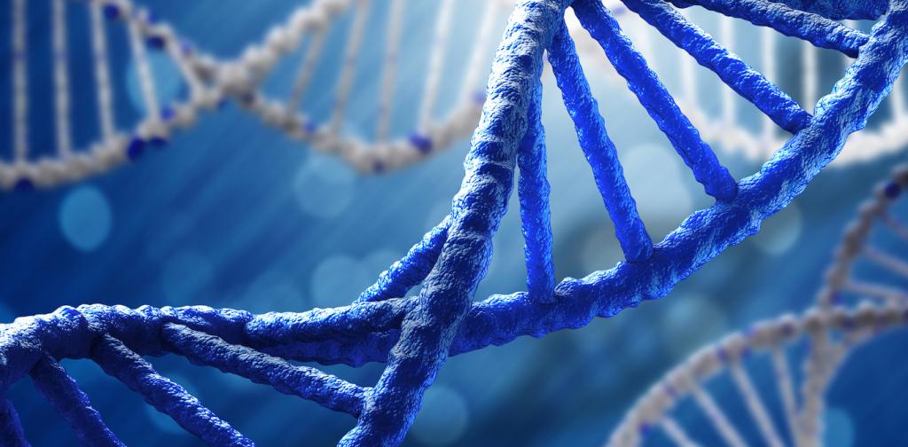
What is the basis of DNA hybridization? Although a double-stranded DNA sequence is generally stable under physiological conditions, changing these conditions in the laboratory (usually by increasing the ambient temperature) will cause the molecules to be separated into separate strands. The latter are complementary to each other, but can also complement other sequences present in their environment. Lowering the ambient temperature allows single-stranded molecules to anneal or “hybridize” with each other. This is the method of DNA hybridization.
Concept from the point of view of molecular biology
Scientists involved in both DNA replication and transcription of DNA into RNA rely on cross-nucleotides and molecular biology methods. This includes Southern blots and Northern blots, polymerase chain reaction (PCR) and most DNA-RNA sequencing and hybridization approaches.
Application
Hybridization is the main property of nucleotide sequences and is used in numerous molecular biology methods. The general genetic relationship of the two species can be determined by hybridizing their DNA segments (DNA-DNA hybridization). Due to the similarity of sequences between closely related organisms, a higher temperature is required to thaw such DNA hybrids compared to more distant organisms. Various methods use hybridization to determine the origin of a DNA sample, including the polymerase chain reaction (PCR). In another method, short DNA sequences hybridize with cellular mRNA to identify expressed genes. Pharmaceutical companies are exploring the use of antisense RNA to bind to unwanted mRNA, preventing the ribosome from converting mRNA to protein.
DNA-DNA hybridization usually refers to a molecular biology method that measures the degree of genetic similarity between pools of DNA sequences. It is commonly used to determine the genetic distance between two organisms. It has been widely used in phylogeny and taxonomy.
Methodology
The DNA of one organism was labeled, then mixed with unlabeled DNA, which could be compared with it. The mixture was incubated to allow the DNA chains to dissociate and then cool to form renewed hybrid double-stranded DNA. Hybridized sequences with a high degree of similarity will bind more tightly and require more energy to separate them: that is, they separate when heated at a higher temperature than dissimilar sequences, a process known as DNA melting.
DNA melting
Assessing the melting profile of hybridized DNA, double-stranded DNA is bound to a so-called “column”, and the resulting mixture is heated. At each stage, the column is washed, and the DNA sequences that melt become single-stranded and wash off the column. The temperatures at which the labeled DNA leaves the column reflects the amount of similarity between the sequences (and the self-folding pattern serves as a control). These results are combined to determine the degree of genetic similarity between organisms. According to modern microbiology, DNA hybridization is impossible without understanding these things.
When several types of ribonucleic (or disoxyribonucleic) acid are compared in this way, similarity values allow the species to be placed in a phylogenetic tree. Therefore, this is one of the possible approaches to conducting molecular taxonomy. Charles Sibley and John Alquist, pioneers of this technique, used DNA-DNA hybridization to study the phylogenetic relationships of birds (Sibley-Alquist taxonomy) and primates.
Importance for Biology
DNA-DNA hybridization is the gold standard for distinguishing bacterial species with a similarity of more than 70%, which indicates that the compared strains belong to different species. In 2014, a threshold of 79% similarity was proposed for the separation of the bacterial subspecies.
Critics argue that the technique is inaccurate for comparing closely related species, since any attempt to measure differences between orthologic sequences between organisms is overloaded with hybridization of paralogous analogs in the body’s genome. DNA sequencing and computational sequence comparisons are currently usually a method for determining genetic distance, although this approach is still used in microbiology to help identify bacteria.
A modern method is to conduct DNA-DNA hybridization in silicone using fully or partially sequenced genomes. Developed by DSMZ, the GGDC is the most accurate known tool for calculating DDH-like values. Among other algorithmic improvements, he solves the problem with paralogous sequences by carefully filtering them from the matches between the two sequences of the genome.
FISH Method
Fluorescence In Situ Hybridization (FISH) is a laboratory method used to detect and determine the DNA sequence, often on a particular chromosome.
In 1969, Joseph Gall and Mary Lou Pardoux published a document demonstrating that radioactive copies of the ribosomal DNA sequence can be used to detect complementary DNA sequences in the frog's nucleus. Starting from these original observations, many refinements have increased the universality and sensitivity of the procedure to such an extent that in situ hybridization (“in place”, Latin) is currently considered an important tool in cytogenetics. (The term in situ now also refers to the initial stage of carcinoma growth, when only epithelial tissue is involved in the pathological process.)
Fluorescence hybridization sequence
RNA probes can be designed for any gene or any sequence within a gene to visualize lncRNA and miRNA mRNA in tissues and cells. FISH is used by studying the cell propagation cycle, in particular the nuclear interphase for any chromosomal abnormality. FISH allows you to analyze a large series of archival cases, it is much easier to identify the identified chromosome, creating a probe with an artificial chromosome base that will attract similar chromosomes.
Hybridization signals for each probe when a nuclear anomaly is detected: each probe for detecting mRNA and lncRNA consists of 20 pairs of oligonucleotides, each pair covers a space of 40-50 b. n. To detect mRNA, probes use proprietary chemistry.
DNA probe hybridization
Probes are often obtained from DNA fragments that have been isolated, purified and amplified for use in the design of the human genome. The size of the human genome is so large compared to the length that can be sequenced directly that it is necessary to divide it into fragments. Ultimately, these fragments were tidied up by digesting a copy of each fragment into even smaller elements using sequence-specific endonuclease to measure the size of each small fragment using size exclusion chromatography using this information to determine where large parts overlap with each other .
To preserve the elements with their individual DNA sequences, fragments were added to the system of constantly repeating bacterial populations. Clonal bacterial populations, each population supporting a single artificial chromosome, are stored in various laboratories around the world. Artificial chromosomes (BACs) can be grown, removed and labeled in any laboratory containing a library. Genomic libraries are often named after the institutions in which they were developed. An example is the RPCI-11 library, named after the Roswell Cancer Institute in Buffalo (New York, USA). These fragments make up about 100 thousand base pairs and are the basis of most FISH probes.