Gram staining is widespread in microbiology, as it is one of the easiest ways to differentiate bacteria depending on the composition of their cell wall. According to Gram, all bacteria can be divided into gram-positive (Gram (+)) and gram-negative (Gram (-)). The Gram stain method was developed in 1884, and since then has not lost popularity, although it has been modified several times.
Cell wall structure
Gram staining reveals whether a particular bacterium is gram-positive or gram-negative. The division of bacteria into Gram (+) and Gram (-) is carried out in accordance with the structure of their cell wall.
The cell wall in the greatest amount contains peptidoglycan (murein) - a complex substance, which includes peptapeptide and glycan. Glycan consists of alternating residues of N-acetylglucosamine and N-acetylmuramic acid, connected to each other by β-1,4-glycosidic bonds. Peptidoglycan provides maintenance of cell shape, osmotic protection, as well as antigenic functions.
The main differences between gram-positive and gram-negative bacteria
In different bacteria, the thickness of the peptidoglycan layer is not the same. In bacteria, which are classified as gram-positive, it is from 15 to 80 nm, while in gram-negative - from 2 to 8 nm. At the same time, gram-negative bacteria under the peptidoglycan layer have a special structure that gram-positive bacteria do not have - the periplasmic space. This space is filled with hydrolytic enzymes - β-lactamase, ribonuclease 1, phosphatase. It is these enzymes that are responsible for the resistance of gram-negative bacteria to many antibiotics.
Layer of peptidoglycan Gram (-) bacteria is associated with a lipopolysaccharide, an antigenic structure containing endotoxin. In Gram (+) bacteria, teichoic acids perform similar functions.
Gram-negative bacteria have an additional structure - the outer membrane.
The essence of the staining method
Before starting staining, prepare smears of the studied bacteria. To do this, water is dripped onto a glass slide and a microorganism culture is added there with a bacterial loop. Then, after the water has completely dried, the smear is fixed - a glass slide is carried several times over the burner flame. Gram stain is more effective than staining live bacteria - dye molecules bind better to dead cells.
Staining is carried out in several stages:
- Small pieces of filter paper are applied to a fixed smear and the main dye is poured - gentian violet or methylene blue.
- After 3-5 minutes, the stained filter paper is removed and the smear is poured with a Lugol solution for 1 minute. In this case, the drug darkens.
- Lugol's solution is drained and the smear is treated with pure ethanol: a few drops are added to the preparation, after 20 seconds it is drained. The procedure is repeated 2-3 times.
- Washed glass with the test drug with distilled water.
- An additional staining is performed - the drug is stained with fuchsin. After 1-2 minutes, the dye is washed off.
- After the water has dried, the smear is examined under a microscope. Gram-positive bacteria will have a blue-violet color, gram-negative - pink or red.
The reasons for the different nature of staining
As described above, when staining bacteria with Gram, gram-positive bacteria stain blue-violet, and gram-negative - in red or pink. The reason for the differential staining of bacteria by this method is that after the soluble form of the gentian violet enters the cell, the dye transforms into an insoluble iodine form. During the treatment of bacteria with ethyl alcohol, lipids are extracted from the membrane by the action of this non-polar solvent. After this, the membrane becomes porous and is no longer a significant obstacle to the washing out of the dye. However, peptidoglycan is more resistant to the action of non-polar solvents, including alcohol. It is it that prevents the dye from being washed out, therefore bacteria with a thick murein layer turn blue-violet (gram-positive) and do not change color after treatment with alcohol.
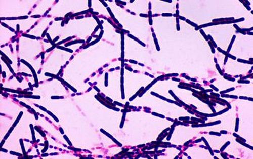
A thin murein layer of gram-negative bacteria cannot retain dye molecules in the cell, therefore, after the action of alcohol, they become colorless - they are painted gram-negative.
After exposure to a smear with fuchsin, when gram stained, gram-positive bacteria remain blue-violet, and gram-negative acquire a pink-red hue.
Examples of Gram (+) and Gram (-) bacteria
Gram-negative include cyanobacteria, sulfur bacteria, iron bacteria, chlamydia, rickettsia, acetic bacteria, many methyl bacteria, thionic bacteria, arsenite bacteria, carboxy bacteria.
Gram-positive are bifidobacteria, many aquatic bacteria, streptococci and staphylococci.