In the process of studying how the brain changed during evolution, an idea was developed about the presence of its three levels. The first of them (the highest) is the anterior section. It includes basal subcortical nodes, cerebral cortex , diencephalic and olfactory brain. The middle section refers to the middle level. And to the lower, there is the posterior part, which consists of the medulla oblongata, cerebellum and pons.
The midbrain, the functions and structure of which we will examine in detail, develops mainly under the influence of the visual receptor during phylogenesis. Consequently, its most important formations relate to the innervation of the eye.
Also, hearing centers were formed in it, later along with the centers of vision, which grew and formed 4 mounds of the roof of the midbrain. We will examine its structure in detail below. And the functions of the midbrain are described in the second half of this article.
Midbrain development
The visual and auditory centers located in it became subcortical, intermediate, having fallen into a subordinate position with the appearance in humans and higher animals of the cortical end of the visual and auditory analyzers in the forebrain cortex. The development of the forebrain in humans and higher mammals has led to the fact that the pathways connecting the cortex of the final to the spinal began to pass through the midbrain, the functions of which have changed somewhat. As a result of this, the latter includes:
- subcortical auditory centers;
- visual subcortical centers, as well as the nuclei of nerves that innervate the muscles of the eye;
- all descending and ascending pathways that connect the cerebral cortex to the spinal cord and pass through the middle transit;
- beams of white matter connecting the midbrain with various parts of the central nervous system.
Structure
The midbrain, the functions and structure of which we are interested in, is the most simply arranged and smallest department (in the photo above it is indicated in brown). It distinguishes the following 2 main parts:
- legs, where conductive paths mainly pass;
- subcortical centers of vision and hearing.
Midbrain roof
The roof of the midbrain, the dorsal part, is hidden under the corpus callosum (its posterior end). It is subdivided into 4 mounds located in pairs by means of two grooves (transverse and longitudinal) running crosswise. The upper two mounds are the subcortical centers of vision, and the lower two are the hearing. Between the upper tubercles in the flat groove is a pineal body. The handle of the knoll is directed laterally, up and anteriorly, to the diencephalon. Every mound passes into it. The handle of the upper mound runs under the thalamus cushion towards the lateral bent body. The lower handle disappears under the cranked medial body. The cranked bodies mentioned above are no longer related to the middle, but to the diencephalon.
Brain legs
We continue to describe the human midbrain, functions and structure. The next thing we stop at is his legs. What is it? This is the ventral part, in which all the pathways leading to the forebrain are located. Note that the legs are two semi-cylindrical thick white strands diverging at an angle from the edge of the bridge and plunging into the hemispheres.
What is the midbrain cavity?
Many terms can be found in a section like midbrain anatomy. The structure and its functions are required in the description of strict scientific accuracy. We omitted complex Latin names, leaving only the basic terms. For the first acquaintance, this is enough.
Let's say a few words about the cavity of the midbrain. It represents a narrow canal and is called a water supply system. This canal is lined with ependyma, it is narrow, its length is 1.5-2 cm. A brain water supply connects the fourth ventricle to the third. The cap of the legs limits it ventrally, and dorsally the roof of the midbrain.
Cross sections of the midbrain
We continue our story. Features of the midbrain of a person can be better understood by looking at it in a cross section. In this case, the following 3 main parts are distinguished in it:
- lid plate;
- tire;
- ventral section, that is, the base of the leg.
Midbrain nuclei
Under the influence of the visual receptor, according to how the midbrain is developed, various nuclei are located in it. The functions of the midbrain nuclei relate to the innervation of the eye. The superior colliculus in the lower vertebrates is the main place where the optic nerve ends, as well as the main visual center. In humans and mammals with the transfer to the forebrain of the visual centers, the connection remaining between the superior mound and the optic nerve is important only for reflexes. In the cranked medial body, as well as in the nucleus of the lower mound, the auditory loop fibers end. The roof of the midbrain is connected to the spinal bilateral connection. The plate of this roof can be considered the reflex center for movements that occur mainly under the influence of auditory and visual irritations.
Brain water supply
He is surrounded by a central gray substance, in its function related to the vegetative system. Under its ventral wall, in the cap of the pedicle of the brain, are the nuclei of two cranial motor nerves.
The core of the oculomotor nerve
It consists of several sections of the innervation of various muscles of the eyeball. Behind and medially from it there is a paired small additional vegetative nucleus, as well as a median unpaired. The unpaired median and additional nucleus innervate the muscles of the eye, which are involuntary. We attribute this part of the oculomotor nerve to the parasympathetic system. Rostrally (above) the nucleus of the oculomotor nerve, the nucleus of the longitudinal medial bundle is located in the cap of the pedicle of the brain.
Brain legs
They are divided into the base of the legs (ventral part) and the tire. Black matter serves as the boundary between them. It owes its color to melanin, the black pigment found in the nerve cells that make it up. The tire of the midbrain is its part located between the black matter and the roof. The central tympanic path leaves from it. This is a descending projection of the nerve pathway, which is located in the lining of the midbrain (its central part). It consists of fibers that go from the red nucleus, the pale ball, the reticular formation of the middle brain and the thalamus to the olive and the reticular formation of the medulla oblongata. This pathway is part of the extrapyramidal system.
Midbrain functions
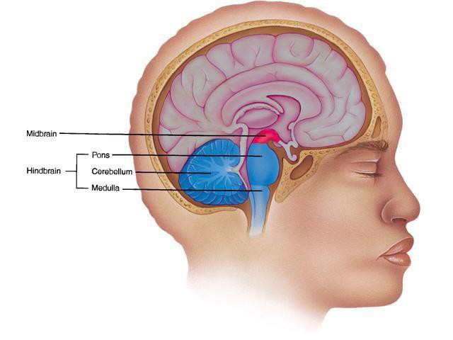
It plays a very important role in the formation of rectifying and installation reflexes that make walking and standing possible. In addition, the midbrain functions as follows: it regulates muscle tone, takes part in its distribution. And this is a necessary condition for the implementation of coordinated movements. Another function - thanks to it, a number of autonomic processes are regulated (swallowing, chewing, breathing, blood pressure). Due to watchdog auditory and visual reflexes, as well as an increase in the tone of the flexor muscles, the midbrain (it is highlighted in red in the photo above) prepares the body to give an answer to the sudden onset of irritation. Statokinetic and static reflexes are realized at its level. Tonic reflexes provide restoration of balance, posture, which was disturbed as a result of a change in position. They appear when the position of the head and body in space changes due to the excitation of proprioreceptors, as well as tactile receptors located on the skin. All these functions of the midbrain indicate that it plays an important role in the body.
Cerebellum
We now turn to the consideration of the cerebellum. What is it? This is the structure of the rhomboid. It is formed in ontogenesis from the cerebral rhomboid bubble (its dorsal wall). It is associated with various parts of the nervous system that control our movements. Its development takes place along the path of improving connections with the spinal cord, as well as weakening them with the vestibular system.
Research by Luigi Luciani
The functions of the midbrain and cerebellum were studied by Luigi Luciani, an Italian physicist. In 1893, he performed experiments on animals with a fully or partially removed cerebellum. He also conducted an analysis of his bioelectric activity, registering it with irritation and at rest.
It turned out that the tone of the extensor muscles increases when half the cerebellum is removed. The limbs of the animal are extended, the body bends, and the head deviates to the operated side. There are movements in a circle ("maneuvrable movement") in the operated direction. The described violations are gradually smoothed out, however, a certain discoordination of movements is maintained.
If you remove the entire cerebellum, severe movement disorders occur. They are smoothed out gradually due to the fact that the cerebral cortex (its motor zone) is activated. However, the animal still remains in violation of coordination. Inaccurate, awkward, sweeping movements, a shaky gait are observed.
Contribution of Academician Orbeli
In 1938, Academician Orbeli discovered that the cerebellum also affects the receptor apparatus and vegetative processes. In addition, there is a relationship with the state of the muscles of the internal organs. Changes in the composition of blood, blood circulation, respiration, digestion, which occur under the influence of the cerebellum, are aimed at providing (trophic) activity of skeletal muscles.
Academician Orbeli considered the cerebellum not only as an assistant to the cerebral cortex in the regulation of muscle movements and tone, but also as an adaptive-trophic center. In this role, it affects all parts of the brain through the nervous system (sympathetic department). This is how metabolism is regulated, and the central nervous system adapts to environmental conditions. It was found that the activity of the cerebellum is inextricably linked with the cerebral cortex and occurs under its control.
Conclusion
So, we briefly examined the cerebellum and midbrain of a person. Their functions have been described by us. Now you know what important role they play. Our body is generally arranged so that all its organs do their job, all of them are necessary. The functions of the medulla oblongata and midbrain, as well as other parts of the body, should be known.
And in conclusion, a few more words. The brain is a complex aggregate of billions of cells working together. He supports life in a flexible and unique but unchanging way and is able to respond to changing incentives, behavioral guidelines and needs. As we move from infancy to childhood, and then to youth, adulthood and old age, throughout the course of our lives, our body follows the same path with us. Accordingly, the brain is changing. It follows, on the one hand, hard-coded evolutionary and ontogenetic patterns of development. But on the other hand, he is able to adapt to a change in the interactions between the external environment and the body.