The skeleton serves as a point of attachment of muscles, is a support for soft tissues, protection and a container of internal organs. It develops from mesenchyme. The human skeleton consists of about two hundred separate bones. The axial skeleton and the additional one are made up of different bones, however, almost all of them with the help of ligaments, joints and other joints form a single whole.
Skeleton changes throughout life
The skeleton is constantly changing throughout life. The cartilaginous skeleton of the fetus, for example, during intrauterine development is gradually replaced by bone. This process continues after birth for several years. A child who has just been born has almost 270 bones in the skeleton. This is much more than that of an adult, in whom it consists of 200-208. This difference arose because the skeleton of the newborn contains many small bones. Only at a certain age they grow together in large ones. This applies, for example, to the bones of the spine, pelvis and skull. The sacral vertebrae fuse into the sacrum (a single bone) only at the age of 18-25 years.
What bones are not directly related to the skeleton?
The skeleton does not directly include six special bones that are in the middle ear, three on each side. They connect only with each other and take part in the work of the organ of hearing. These bones transmit vibrations to the inner ear from the eardrum.
Features of some bones
The hyoid bone in the human body is the only one not directly related to others. It is located on the neck, but it is traditionally attributed to the bones of the skull (facial section). She is suspended from him by muscles and connected to the larynx. The femur is the longest in the skeleton, and the stirrup located in the middle ear is the smallest.
Skeleton organization
In humans, the skeleton is arranged according to the principle common to vertebrates. Its bones are divided into the following two groups: axial and additional skeleton. The first is the bones that form the skeleton of the body. They lie in the middle - these are all bones of the neck and head, sternum, ribs, and spine. The axial skeleton of animals is built on the same principle. Additional - this is the scapula, collarbone, bones of the upper and lower extremities and pelvis.
Axial skeleton bone subgroups
All bones of the skeleton are divided into subgroups. The axial skeleton consists of the following.
1. The skull is the bone base of the head, as well as the receptacle of the brain, olfactory organs, hearing and vision. It distinguishes two departments: facial and brain.
2. Considering the human skeleton (axial skeleton), one should also note the chest, which is a compressed truncated cone in shape. It is a receptacle for various internal organs. It consists of 12 pairs of ribs, 12 thoracic vertebrae, as well as the sternum.
3. The spine (otherwise - the spinal column) is the support of the entire skeleton, the main axis of the body. The spinal cord passes inside the spinal canal.
Subgroups of bones of the extra skeleton
The following subgroups are distinguished in it.
1. The belt of the upper extremities, which provides accession to the axial skeleton of the upper limbs. It consists of paired clavicles and shoulder blades.
2. The upper limbs, which are most suited for the implementation of labor activity. They consist of three sections: hand, forearm and shoulder.
3. Belt of the lower extremities, which provides accession to the axial skeleton of the lower extremities. In addition, it is the support and receptacle for the organs of the genital, urinary and digestive systems.
4. The lower limbs, which provide movement of the human body in space.
Bones and sections of the axial skeleton
As you can see, skeleton bones belong to two groups. We briefly reviewed the axial and additional skeleton. On the additional, we will not dwell in detail, since this is not part of our task. Now consider the various departments and bones that make up the axial skeleton.
Vertebral column
This is the mechanical support of the body. It consists of 32 to 34 vertebrae, interconnected. Five departments are allocated in a backbone: coccygeal, sacral, lumbar, thoracic, cervical. Connections in the lumbar and cervical regions are mobile, and in the sacral and thoracic - inactive. Four physiological bends has a vertebral column. The lumbar and cervical bend is directed forward, forming lordosis, and the sacral and thoracic - backward (kyphosis). In different departments, the size of the vertebrae varies. They depend on the size of the load falling on one or another of them and on the development of muscles. The sacral and lumbar vertebrae reach their maximum size. Intervertebral discs act as a shock absorber - distribute pressure between different vertebrae, and also provide the necessary strength and mobility.
During life, the axial skeleton develops. In a newborn, the spinal column is almost straight, after some time, bends of the spine appear. There are two bends back and two forward (kyphosis and lordosis).
Their main purpose is to weaken the concussion of the torso and head when running, walking, jumping. Scoliosis (curvature of the spine in any direction) is observed in many people. Often it is a consequence of painful changes occurring in the spine.
Vertebrae
The axial skeleton includes vertebrae. They have a round body, as well as an arc closing the vertebral foramen. They have processes that connect the articulating vertebrae. The spinal cord passes inside all openings. The tunnel they formed is called the spinal canal. This is a reliable bone protection for the spinal cord located in it. The composition of the vertebra includes: dura mater (protective membrane); spine-shaped bone process that connects it to the muscles; spinal cord and blood vessels. In the section of the intervertebral disc, a biconvex gelatinous nucleus and fibrous rings can be seen. The spinous process is turned back, and the vertebral body is forward. In the middle is the vertebral foramen. Let's say a few words about arcs. On the arches of the vertebrae there are recesses that together form the intervertebral foramen through which the spinal nerves pass.
Let us dwell in more detail on some vertebrae, considering the structure of the axial skeleton. Atlas is the first cervical vertebra. He has no body. This vertebra articulates with the 2nd cervical vertebra and with the occipital bone of the skull. The epistrophy (2nd cervical vertebra) has a tooth-like process that connects to the atlas (its anterior arch). The spinous process of the 7th cervical vertebra is not bifurcated. It is easily palpated. This process protrudes above neighboring vertebrae, their spinous processes. It is more noticeable in men. On the thoracic vertebrae are articular fossae. They are needed to attach the ribs. The spinous processes in the thoracic vertebrae are directed down and back, they are the longest. The most massive are the lumbar vertebrae. Their spinous processes lean back. The sacrum consists of 5 fused vertebrae. There are a wide upper part (base), two lateral parts and a lower narrow one (apex). Nerves pass through the holes in the sacrum, and the sacral canal is located inside. It is a continuation of the spinal canal. The pelvis is attached to the sacrum. The coccygeal bone of the axial skeleton is divided into 4-5 underdeveloped vertebrae fused together. These are the remains of the tail that the ancestors of man had. With the help of joints, cartilage and ligaments, the vertebrae are connected. The spine can bend and bend, twist, bend to the side. Its most mobile departments are cervical and lumbar.
Rib cage
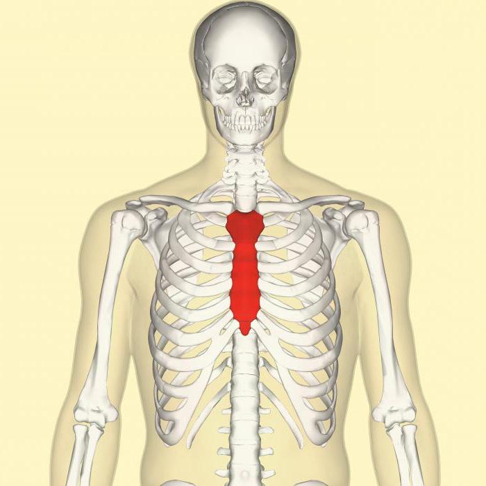
Another section that has an axial skeleton is the chest. It consists of a sternum (highlighted in red in the photo), ribs and thoracic vertebrae. The length of the sternum in adults is from 16 to 23 cm. This is an unpaired flat bone of the axial skeleton. The following three parts are distinguished in it: the xiphoid process, the middle (body) and the upper (handle). The ribs are made up of cartilage and bone. The first of them is located almost horizontally. Seven pairs of ribs with their cartilage in the front ends connect to the sternum. The other five pairs do not connect with her. The 8th, 9th and 10th pair are attached to the cartilage of the overlying rib. The 11th and 12th freely end with the front ends in the muscles. In humans, the chest contains the lungs, heart, esophagus, trachea, nerves and large vessels. She participates in breathing - her volume during exhalation and inhalation due to rhythmic movements decreases and increases. In a newborn, the chest has a pyramidal shape. However, it changes with the growth of the chest. In women, it is smaller than in men, and their upper part is relatively wider in them. A change in the chest is possible after diseases. For example, chicken breast develops with severe rickets (in this case, the sternum comes forward sharply).
Skull bones
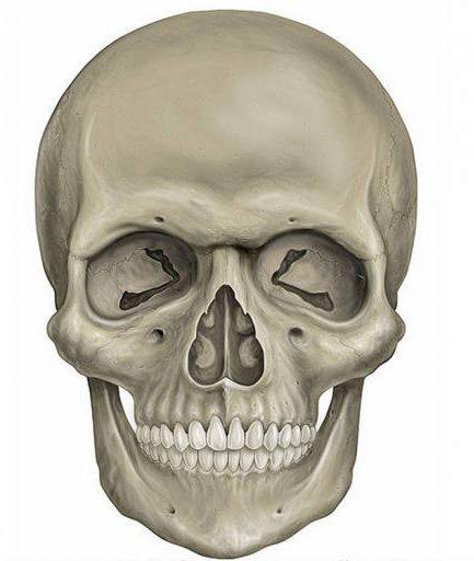
Describing the axial skeleton, you need to talk about the skull. Its bones consist of the following parts: nasal bone, frontal bone, parietal, zygomatic, occipital, lower and maxillary bones and teeth. The skull (skeleton of the head) has a cavity where the brain is located. There are, in addition, the oral cavity, nose, receptacle for the organs of hearing and vision. Examining the axial skeleton of animals and humans, the facial and brain parts of the skull are usually distinguished. All of his bones, except the lower jaw, are connected by seams. Two paired bones make up the brain. We are talking about the temporal and parietal. It also distinguishes 4 unpaired - occipital, ethmoid, wedge-shaped, frontal. Six paired bones represent the front section (upper jaw, lacrimal, nasal, palatine, zygomatic and lower nasal concha), as well as two unpaired. The latter include the opener and lower jaw. The hyoid bone is also the face bone. Many bones of the skeleton of the head have channels and openings for the passage of blood vessels and nerves. Some of them have cells or cavities filled with air (they are called sinuses). The brain part of the skull in humans prevails over the facial.
Sutures of the cranial bones
The seams that connect the bones of the skull are different. They are flat (the edges of the bones are adjacent to each other with even edges), scaly (the parietal and temporal bones are connected this way), and dentate (they are characteristic of the main part of the bones of the skull and are the strongest). Most sutures in adults and especially in older people are ossified. Using the temporomandibular combined joint, the lower jaw is connected to the temporal bones. There is cartilage in this joint, the joint capsule is strengthened by ligaments.
More on the structure of the skull
The roof is the upper part of the cerebral skeleton of the head. And the bottom is the foundation. It has a large occipital foramen. The facial part of the bone (with the exception of the lower shell), as well as the roof of the skull, undergo 2 stages in their development: first, membranous, then bone. For the other bones of the skull, three stages are characteristic: membranous, cartilage and bone. The remains of the membranous skull (they are called fontanelles) are in the roof of the skull of the newborn. There are only six of them: two mastoid, two wedge-shaped, posterior and anterior. The largest of them are rear and front. The anterior one is at the junction of the parietal and frontal bones (at the crown). By one and a half years, it ossifies. The occipital (posterior) fontanel overgrows 2 months after the birth of the baby. In full-term children, lateral fontanels are usually absent, and if they are, they also quickly overgrow (at the 2nd or 3rd month of life). In the newborn, the facial section is less developed than the brain than in the adult: teeth are missing, the airways of the cranial bones are not developed. The sutures are ossified by old age, and the layer of spongy substance also decreases in the bones - the skull becomes fragile and light. Its growth is completed by 25-30 years. The skull of men is relatively larger than women, which is associated with overall body size. Mounds and protrusions on the cranial bones are less pronounced in women than in men.
So, we examined the main sections of the axial skeleton. Recall that we talked about the incremental only briefly, since it is not the subject of this article. Now you know that the axial skeleton consists of various bones, which have different structures and functions.