The spindle of division is a temporary structure that forms during the processes of mitosis and meiosis, and ensures the segregation of chromosomes and cell division. It is bipolar: the microtubule system formed in the space between the poles resembles a spindle in shape. In the centromere region, spindle microtubules join the kinetochores of the chromosome. On them, the chromosomes move to the poles.
Structure
The division spindle consists of three main structural elements: microtubules, division poles, and chromosomes. The division poles in animals are organized using centrosomes, which contain centrioles. In the absence of a centrosome (in plants, and in oocytes in some animal species), the spindle has wide poles and is called acentrosomal. Another structure is involved in spindle formation - motor proteins. They belong to dyneins and kinesins.
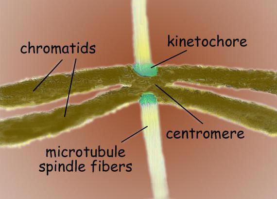
The spindle of division is a bipolar structure. At both poles are centrosomes - organelles, which are the centers of microtubule organization. In the structure of the centrosome, two centrioles are distinguished, surrounded by many different proteins. Condensed chromosomes in the form of two chromatids bonded to a centromere site are located between the poles. In the region of the centromeres there are kinetochores, to which microtubules attach.
Formation
Since the division spindle is the structure responsible for cell division, the beginning of its assembly occurs in prophase. In plants and in oocytes, in the absence of centrosomes, the core membrane is the center of microtubule organization. Microtubules approach the nuclear envelope and at the end of the prophase their orientation ends, and a “prophase spindle” is formed - the axis of the future division spindle.
Due to the fact that it is the centrosome that acts as the center of organization in animal cells, the beginning of the formation of the spindle of division is the divergence of the two centrosomes during the prophase. This is possible due to motor proteins dyneins: they attach to the outer surface of the nucleus, as well as to the inner side of the cell membrane. The group of dyneins attached to the membrane is connected to the astral microtubules and they begin to move towards the minus end, due to which the centrosome is diluted in opposite parts of the cell membrane.
End of assembly
The final formation of the fission spindle occurs at the prometaphase stage, after the disappearance of the core membrane, it becomes full, because it is after this that the centrosomes and microtubules can gain access to the spindle components.
However, there is one exception: in budding yeast, the formation of a fission spindle occurs inside the nucleus.
The formation of fission spindle threads and their orientation is impossible without two processes: the organization of microtubules around the chromosomes and their attachment to each other at opposite fission poles. Many elements necessary for the final formation of the fission spindle, including chromosomes and motor proteins, are located inside the cell nucleus, and microtubules and, if it is an animal cell, centrosomes are contained in the cytoplasm, that is, the components are isolated from each other. That is why the formation of the spindle ends only after the disappearance of the nuclear shell.
Chromosome Accession
Protein, as well as many other structures, are involved in the formation of the fission spindle, and this process has been well studied in animal cells. During the prophase period, microtubules form a star-shaped structure around the centrosome, which diverges in the radial direction. After the core membrane is destroyed, dynamically unstable microtubules begin to actively probe this region and kinetochores of chromosomes can be fixed on them. Some part of the chromosomes immediately appears at opposite poles, while the rest are first associated with microtubules of one of the poles, and only then they begin to move towards the desired pole. When the process is completed, the chromosomes already connected to a pole begin to be attached by kinetochores to microtubules from the opposite pole, thus, during the metaphase process, ten to forty tubes are attached to kinetochores. This formation is called a kinetochore beam. Gradually, each of the chromosomes becomes associated with the opposite pole, and they form a metaphase plate in the central part of the fission spindle.
Second option
There is another scenario in which a spindle of division can form. This is possible both for cells with centrosomes and for cells in which they are absent. The gamma-tubulin ring complex is involved in the process, due to which there is nucleation of short microtubules around the chromosomes. The tubules are attached to the kinetochores with the plus-end, after which the polymerization of microtubules begins, that is, regulated growth. The minus ends "merge" and remain at the fission poles due to motor proteins. If a centrosome pair is involved in the formation of the fission spindle, this facilitates the connection of microtubules, but the process is possible without them.
Equally
A clear separation of chromosomes between two cells formed during division can occur only if paired chromatids with their kinetochores joined at different poles. The bipolar discrepancy between chromatids is called amphithepic, but there are other options that arise during the division of the spindle. This is monothepic (one kinetochore joins one pole) and syntepic (both kinetochore chromosomes connect to one pole). In case of a merotepic one kinetochore is captured at once by two poles. Stable is only the usual, bipolar bonding, which occurs due to the pulling forces from the poles, other bonding methods are unstable and reversible, but possible due to the location of the kinetochore.