The brain is an organ that regulates all body functions. It is included in the central nervous system. Leading scientists and physicians from different countries have been studying and continue to study the brain.
General information
The brain includes 25 billion neurons that make up gray matter. Body weight varies by gender. For example, in men its weight is about 1375 g, in women - 1245 g. On average, its share in total body weight is 2%. At the same time, scientists have established that the level of intellectual development is not connected with the mass of the brain. Mental abilities are affected by the number of connections created by the body. Brain cells are neurons and glia. The former generate and transmit impulses, the latter perform additional functions. There are cavities inside the brain. They are called ventricles. In different parts of the human body, cranial nerves depart from the organ we are examining. They are paired. In total, 12 pairs of nerves depart from the brain. Three membranes cover the brain: soft, hard and arachnoid. Between them there are spaces. They circulate cerebrospinal fluid. It acts as an external hydrostatic environment for the central nervous system, and also provides the excretion of metabolic products. Shells of the brain differ in their structure and the number of vessels passing through them. However, they all protect the contents of the upper part of the skull from mechanical damage.
Spider Web MO
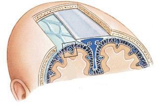
Arachnoidea encephali is separated from the hard shell by the capillary network of the subdural space. It does not go into the grooves and furrows, like a vascular. However, the arachnoid membrane is thrown through them in the form of bridges. As a result, a subarachnoid space forms, which fills a clear fluid. In some areas, mainly on the basis of the brain, a particularly good development of subarachnoid spaces is noted. They form deep and wide containers - tanks. They contain cerebrospinal fluid.
Vascular (soft) MO
Pia mater encephali directly covers the brain surface. It is presented in the form of a transparent two-layer plate, which extends into the crevices and grooves. In vascular MO, there are chromatophores - pigment cells. Especially a lot of them are revealed on the basis of the brain. In addition, there are lymphoid, mast cells, fibroblasts, numerous nerve fibers and their receptors. Parts of soft MO accompany arterial vessels (medium and large), reaching arterioles. Between their walls and the shell are the spaces of Virchow - Robin. They are filled with cerebrospinal fluid and are in communication with the subarachnoid space. Elastic and collagen fibrils are thrown through them. Vessels are suspended on them, by means of which conditions are created for their displacement during pulsation without affecting the brain substance.
TMO
It is characterized by special strength and density. Its composition contains a large number of elastic and collagen fibers. A hard shell is formed by a dense connective tissue.
Features
The hard shell lining the cavity of the skull from the inside. Along with this, she acts as his inner periosteum. In the area of the large opening in the occipital part, the TMT passes into the hard shell of the spinal cord. It also forms perineural vaginas for cranial nerves. Penetrating into the holes, the shell fuses with their edges. The connection with the bones of the arch is fragile. The shell is easily separated from them. This determines the possibility of epidural hematomas. In the area of the cranial base, the membrane fuses with the bones. In particular, a strong fusion is observed in the areas of connections of the elements with each other and the exit of cranial nerves from the cavity. The inner surface of the membrane is lined with endothelium. This determines its smoothness and pearlescent shade. In some areas, cleavage of the shell is noted. Here its processes are formed. They protrude deeply into the gaps that separate parts of the brain. Triangular channels are formed in the areas where the processes are discharged, as well as in the places of attachment to the bones of the internal cranial base. They are also covered with endothelium. These channels are the sinuses of the dura mater.
Sickle
It is considered the largest process of the membrane. The sickle penetrates the longitudinal gap between the left and right hemispheres, not reaching the corpus callosum. It is a thin sickle-curved plate in the form of 2 sheets. In the split base of the appendix lies the upper sagittal sinus. The opposite edge of the sickle has a thickening also with two petals. The lower sagittal sinus lies in them.
Connection with cerebellum elements
In the anterior part, the sickle is fused with the cockscomb on the ethmoid bone. The posterior region of the appendix at the level of the occipital inner protrusion is connected to the cerebellum. He, in turn, hangs over the cranial fossa with a gable tent. The cerebellum lies in it. His breech penetrates the transverse gap in the large brain. Here it separates the cerebellar hemispheres from the occipital lobes. Roughnesses are present on the front edge of the bastard. Here, a notch forms, to which the cerebral trunk adjoins in front. The lateral patches of the tent fuse with the edges of the groove in the posterior sections on the transverse sinus of the occipital bone and with the upper edges of the pyramids on the temporal bones. The connection extends to the posterior processes of the wedge-shaped element in the anterior parts on each side. In the sagittal plane is the cerebellar sickle. Its cutting edge is free. He shares the cerebellar hemispheres. The back of the sickle is located along the occipital inner crest. It extends to the edge of the large hole and covers it with two legs on both sides. The occipital sinus is present at the base of the sickle.
Other items
A diaphragm stands out in the Turkish saddle. It is a plate located horizontally. In its center is a hole. The plate is stretched over the pituitary fossa and forms its roof. Under the diaphragm is the pituitary gland. It connects through the hole to the hypothalamus with the help of a funnel and stem. In the area of trigeminal depression near the top of the temporal bone, the dura mater diverges by 2 leaves. They form the cavity in which the nerve (trigeminal) node is located.
Dura mater
They are sinuses formed due to the splitting of TMT into two leaves. The sinuses of the brain act as original vessels. Their walls are formed by plates. Sinuses and veins of the brain have a common symptom. Their inner surface is lined with endothelium. Meanwhile, the sinuses of the brain and blood vessels directly differ in the structure of the walls. In the latter, they are elastic and include three layers. When cut, the lumen of the veins decreases. The walls of the sinuses, in turn, are tightly stretched. They are formed by a dense fibrous connective tissue in which elastic fibers are present. When cut, the lumen of the sinuses gapes. In addition, valves are present in the venous vessels. In the sinus cavity there are several incomplete beams and wavy beams. They are covered with endothelium and are transferred from wall to wall. In some sinuses, these elements are characterized by significant development. There are no muscle elements in the walls of the sinuses. The sinuses of the dura mater have a structure that allows blood to flow freely under the influence of its gravity, regardless of fluctuations in intracranial pressure.
Kinds
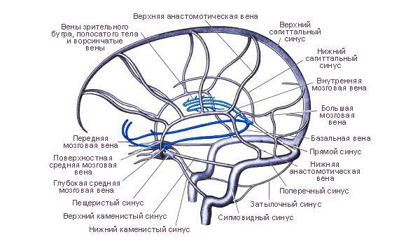
The following sinuses of the dura mater are distinguished:
- Sinus sagittalis superior. The superior sagittal sinus runs along the upper edge of the large sickle, from the cocks to the occipital inner protrusion.
- Sinus sagittalis inferior. The lower sagittal sinus is located in the thickness of the free edge of the large sickle. It flows into the sinus rectus with the back. The joint is located on the site where the lower edge of the large sickle fuses with the front edge of the cerebellar tent.
- Sinus rectus. The direct sinus is located in the splitting of the tent along the line of attachment of a large sickle to it.
- Sinus transversus. The transverse sinus is located at the place where the cerebellum is projected from the lining of the brain.
- Sinus occipitalis. The occipital sinus lies at the base of the cerebellar sickle.
- Sinus sigmoideus. The sigmoid sinus is located in the groove of the same name on the inner cranial surface. It looks like the letter S. In the area of the jugular opening, the sinus passes into the internal vein.
- Sinus cavernosus. The paired cavernous sinus is located on both sides of the Turkish saddle.
- Sinus sphenoparietalis. The sphenoid-parietal sinus is adjacent to the posterior free area on the small wing of the sphenoid bone.
- Sinus petrosus superior. The upper stony sinus is located at the upper edge of the temporal bone.
- Sinus petrosus inferior. The lower stony sinus is located between the occipital slope and the temporal bone pyramid.
Sinus sagittalis superior
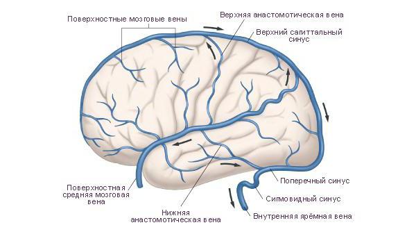
In the anterior sections, the superior sinus anastomoses (connects) with the veins of the nasal cavity. The back runs into the transverse sinus. To the left and to the right of it are side gaps communicating with it. They are small cavities located between the outer and inner sheets of TMT. Their number and size are very different. Lacunae communicate with the sinus sagittalis superior cavity. They include the vessels of the hard shell and brain, as well as diploic veins.
Sinus rectus
The direct sinus acts as a peculiar continuation of the sinus sagittalis inferior from behind. It connects the backs of the sinuses superior and inferior. In addition to the superior sinus, a large vein enters the anterior end of sinus rectus. The back sinus flows into the middle part of the sinus transversus. This area is called the sinus drain.
Sinus transversus
This sinus is the largest and widest. On the inside of the bones of the back of the head it corresponds to a wide furrow. Next, sinus transversus passes into the sigmoid sinus. Then he leaves at the mouth of the internal jugular vessel. Sinus transversus and Sinus sigmoideus, thus, act as the main venous collectors. At the same time, all other sinuses fall into the first. Some venous sinuses enter it directly, some indirectly. Right and left transverse sinus continues in sinus sigmoideus of the corresponding side. The area where the venous sinuses sagittalis, rectus and occipitalis flow into it is called the drain.
Sinus cavernosus
Its other name is the cavernous sinus. He received this name in connection with the presence of numerous partitions. They give the sinus an appropriate structure. Through the cavernous sinus pass the abducent, ophthalmic, block, oculomotor nerve, as well as the carotid artery (internal) together with the sympathetic plexus. There is a message between the right and left sinuses. It is presented in the form of the posterior and anterior interventricular sinus. As a result, a vascular ring is formed in the region of the Turkish saddle . Sinus sphenoparietalis flows into the cavernous sinus (in its anterior sections).
Sinus petrosus inferior
It enters the superior bulb of the jugular (internal) vein. The maze vessels are also suitable for sinus petrosus inferior. The stony sinuses of the dura mater are connected by several vascular channels. On the basilar surface of the occipital bone, they form the plexus of the same name. It is formed by the fusion of the venous branches of the right and left sinus petrosus inferior. Through the occipital foramen, the basilar and internal vertebral vascular plexus are connected.
Additionally
In some areas, the sinuses of the membrane form anastomoses with external venous vessels of the head with the help of graduates - emissary veins. In addition, the sinuses communicate with diploic branches. These veins are located in the spongy substance in the bones of the cranial vault and flow into the superficial vessels of the head. Blood, therefore, flows through the vascular branches into the sinuses of the TMT. Then it flows into the left and right jugular (internal) veins. Due to sinus anastomoses with diploic vessels, graduates and plexuses, blood can flow into the surface networks of the face.
Vessels
The meningeal (middle) artery (branch of the maxillary) approaches the hard membrane through the left and right spinous openings. In the temporal-parietal area of the TMT, it branches. The membrane of the anterior fossa of the skull is supplied with blood from the anterior artery (ethmoid branch of the eye vessel system). In the TMT of the posterior fossa of the skull, the meningeal posterior, vertebral branches and the mastoid branch of the occipital artery branch.
Nerves
The hard shell is innervated by various branches. In particular, branches of the vagus and trigeminal nerves are suitable for it. In addition, sympathetic fibers provide innervation. They enter the hard shell in the thickness of the outer wall of blood vessels. In the area of the cranial anterior fossa, the TMT receives processes from the optic nerve. Its branch - tentorial - provides innervation of the cerebellar outline and the sickle of the brain. The cranial middle fossa is supplied by the meningeal process of the maxillary and part of the mandibular nerves. Most branches run along the vessels of the membrane. In the cerebellum, however, the situation is somewhat different. There are few vessels, and the branches of nerves are located in it independently of them.