With careful consideration, the structure of the hand, like any other department of our musculoskeletal system, is quite complex. It consists of three main structures: bones, muscles and ligaments that hold bones. The brush has three sections, namely: the wrist, fingers and metacarpus.
In this article we will consider in detail the brush: structure, muscles, joints of the hand. Let's start with a description of the bones in its various departments.
Wrist bones
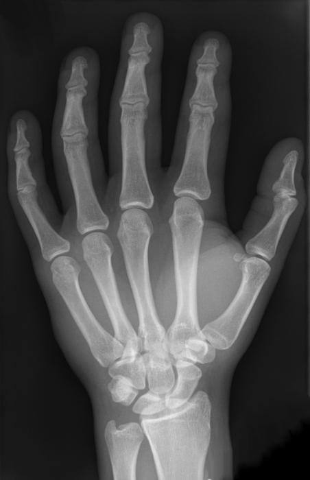
Since the hands must perform fairly precise and intricate movements, the structure of the bones of the hand is also extremely complex. In the wrist - 8 small bones of irregular shape, arranged in two rows. In the figure below you can see the structure of the right hand.
The proximal row forms the articular surface convex to the radius. It includes bones, counting from the fifth to the thumb: pea-shaped, trihedral, lunate and scaphoid. The next row is distal. It connects to the proximal joint of an irregular shape. The distal row consists of four bones: trapezoid, polygonal, capitate and hook-shaped.
Metacarpal bones
This department, consisting of 5 tubular metacarpal bones, also demonstrates the intricate structure of the hand. The skeleton of these tubular bones is complex. Each of them has a body, a base and a head. The metacarpal bone of the 1st finger is shorter than the others and is massive. The second metacarpal bone is the longest. The rest decrease in length with distance from the first and approaching the elbow edge. The bases of the above metacarpal bones are articulated with the bones that form the wrist. The first and fifth metacarpals have bases with articular surfaces of a saddle shape, the others are flat. The heads of the metacarpal bones, having the articular surface (hemispherical), articulate with the proximal digital phalanges.
Finger bones
Each finger, with the exception of the first, which consists of only two phalanges and has no middle, has 3 phalanges: distal, proximal and middle (intermediate). The shortest are distal; proximal - the longest. There is a phalanx head at the distal end, and its base at the proximal end.
Sesamoid bones
In the thickness of the tendons, in addition to these bones, there are sesamoid located between the proximal phalanx of the thumb and its metacarpal bone. There are still inconsistent sesamoid bones. They are located between the proximal phalanges of the fifth and second fingers and their metacarpal bones. Usually sesamoid bones are located on the palmar surface. But sometimes they can be found on the back. The pisiform bone also belongs to the aforementioned species. Sesamoid bones and their processes increase the shoulder strength of the muscles attached to them.
We examined the structure of the hand and the bones of the hand, we now turn to the ligamentous apparatus.
Wrist joint
It consists of the radius and proximal bones of the wrist: trihedral, lunate and scaphoid. The ulna is supplemented by the articular disc and does not reach the wrist joint. The main role in the formation of the elbow joint is played by the ulnar bone. Whereas the wrist band is radial. The wrist joint is ellipsoidal in shape. In it, abduction, adduction of the hand, flexion and extension are possible. A small passive rotational movement (10-12 degrees) is also possible in this joint, but is due to the elasticity of the articular cartilage. Through the soft tissues, it is easy to detect the gap of the wrist joint, which is felt from the elbow and radial sides. With the ulnar, you can feel the groove between the trihedral bone and the head of the ulnar bone. On the radial side there is a gap between the scaphoid and the lateral styloid process.
The movements of the wrist joint are closely related to the work of the mid-carpal joint located between the distal and proximal rows. Its surface is complex, irregular in shape. When bending and unbending, the volume of mobility reaches 85 degrees. The adduction of the hand in the aforementioned joint reaches 40 degrees, the abduction - 20. The wrist joint can circulate, i.e. circular motion.
This joint is strengthened by numerous ligaments. They are located between individual bones, as well as on the lateral, medial, dorsal and palmar surfaces of the wrist. Collateral ligaments (radial and ulnar) play the most important role. On the ulnar and radial sides between the bony elevations is a flexor retainer - a special ligament. In fact, it does not apply to the joints of the hand, as a thickening of the fascia. The flexor retainer converts the furrow of the wrist into a canal in which the median nerve and tendons of the flexors of the fingers pass. We continue to describe the anatomical structure of the hand.
Metacarpal joints
They are flat in shape, inactive. The exception is the thumb joint. The range of motion of the carpal-metacarpal joints is not more than 5-10 degrees. In them mobility is limited, because ligaments are well developed. Located on the palmar surface, they form a stable palmar ligamentous apparatus connecting the wrist and metacarpal bones. There are arched ligaments on the hand, as well as transverse and radial. The capitate is central in the ligamentous apparatus; a large number of ligaments are attached to it. Palms are much better developed than the back. The back ligaments connect the bones of the wrist. They form thickened capsules that cover the joints between these bones. Interosseous are located in the second row of the bones of the wrist.
In the thumb, the carpal-metacarpal joint is formed by the base of the first metacarpal and polygonal bone. Joint surfaces have a saddle shape. This joint can perform the following actions: abduction, adduction, reposition (reverse movement), opposition (opposition) and circumduction (circular motion). The volume of grasping movements, due to the fact that the thumb is opposed to all others, increases significantly. 45-60 degrees is the mobility of the wrist-metacarpal joint of this finger during adduction and abduction, and in the opposite movement and opposition - 35-40.
Hand structure: metacarpophalangeal joints
The named joints of the hand are formed by the heads of the metacarpal bones with the participation of the bases of the proximal phalanges of the fingers. They are spherical in shape, have 3 axes of rotation perpendicular to each other, around which there is an extension and bending, abduction and reduction, as well as circular movements (circumduction). Reduction and abduction is possible at 45-50 degrees, and flexion and extension - 90-100. These joints have collateral ligaments located on the sides that strengthen them. The palmar, or accessory, are located on the palmar side of the capsule. Their fibers are intertwined with the fibers of the deep transverse ligament, which prevents the divergence in different directions of the heads of the metacarpal bones.
Interphalangeal joints of the hand
They are block-shaped, and the axis of their rotation are transverse. Extending and bending is possible around these axes. The proximal interphalangeal joints have a flexion and extension volume equal to 110-120 degrees, distal joints - 80-90. Interphalangeal joints are very well strengthened thanks to the collateral ligaments.
Synovial as well as fibrous sheaths of the tendons of the fingers
The extensor retainer, as well as the flexor retainer, plays a huge role in strengthening the position of the muscle tendons passing under them. This is especially true when the brush is working: when it is unbent and bent. Nature conceived a very competent structure of the hand. Tendons find support in the aforementioned ligaments from their inner surface. The separation of tendons from the bones prevents the ligaments. This allows you to withstand heavy pressure during intense work and strong muscle contraction.
Special tendon sheaths, which are bone-fibrous or fibrous channels, contribute to the reduction of friction and the sliding of tendons going to the wrist from the forearm. They have synovial vaginas. The largest number of them (6-7) is under the extensor retainer. The radius and ulna have grooves that correspond to the location of the tendons of the muscles. And also the so-called fibrotic jumpers, which separate the channels from each other and pass to the bones from the extensor retainer.
Palmar synovial vaginas belong to the flexor tendons of the fingers and hands. The common synovial vagina extends to the center of the palm and reaches the distal phalanx of the fifth finger. Here are the tendons of the superficial and deep flexors of the fingers. The thumb has a long flexor tendon located separately in the synovial vagina and passing to the finger along with the tendon. The synovial vagina in the palm of the hand is devoid of muscle tendons that go to the fourth, second and third fingers. Only the tendon of the fifth finger has a synovial vagina, which is a continuation of the general.
Muscle brush
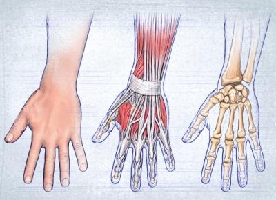
In the picture below you can see the muscles of the arm. The structure of the hand is shown in more detail here.
The muscles in the hand are only on the palmar side. They are divided into three groups: middle, thumb and thumb.
Since finger movements require great accuracy, a significant number of short muscles are located in the hand, which complicates the structure of the hand. The arm muscles of each of the groups will be considered below.
Middle muscle group
It is formed by the vermiform muscles starting from the tendons of the deep flexor of the fingers and attached to the proximal phalanges, more precisely their bases, from the second to fifth fingers, if we consider the structure of the arm. These arm muscles also extend from the dorsal and palmar interosseous, located between the metacarpals, attached to the base of the proximal phalanges. The function of this group is that these muscles are involved in the flexion of the proximal phalanges of these fingers. Thanks to the palmar interosseous muscles, it is possible to bring the fingers to the middle finger of the hand. With the help of the rear interosseous, they are diluted to the sides.
Thumb muscles
This group forms the elevation of the thumb. These muscles begin near the nearby bones of the metacarpus and wrist. As for the thumb, its short flexor is attached near the sesamoid bone, which is located near the base of the proximal phalanx. The opposing thumb muscle goes to the first metacarpal bone, and the adducting thumb is located on the side of the internal sesamoid bone.
Thumb muscles
This muscle group forms an elevation on the inside of the palm of the hand. These include: the muscle that diverts the little finger, the opposing little finger, the short palmar, and also the short flexor.
They originate from nearby bones in the wrist. These muscles are attached to the base of the fifth finger, more precisely its proximal phalanx, and to the fifth metacarpal bone. Their function is reflected in the name.
In the article we tried to most accurately represent the structure of the hand. Anatomy is a fundamental science, requiring, of course, a more thorough study. Therefore, some issues remained unlit. The structure of the hand and wrist is a topic that interests not only doctors. Her knowledge is also necessary for athletes, fitness instructors, students and other categories of people. The structure of the hand, as you noticed, is quite complicated, and you can study it for quite a long time, relying on various sources.