The human nervous system implements complex analytical and synthetic processes that ensure the rapid adaptation of organs and systems to changes in the external and internal environment. The perception of stimuli from the outside world is due to the structure, which includes the processes of afferent neurons containing glial oligodendrocyte cells, or lemmocytes. They turn external or internal stimuli into bioelectric phenomena called excitations or nerve impulses. Such structures are called receptors. In this article, we will study the structure and functions of receptors of various sensory systems in humans.
Types of nerve endings
In anatomy there are several systems for their classification. The most common divides the receptors into simple ones (they consist of processes of one neuron) and complex ones (a group of neurocytes and auxiliary glia cells in a highly specialized sensory organ). Based on the structure of sensory processes. they are divided into primary and secondary endings of a centripetal neurocyte. These include various skin receptors: nociceptors, mechanoreceptors, baroreceptors, thermoreceptors, as well as nerve processes that innervate internal organs. Secondary are derivatives of the epithelium, creating a potential action in response to irritation (receptors of taste, hearing, balance). The rods and cones of the photosensitive membrane of the eye - the retina - occupy an intermediate position between the primary and secondary sensitive nerve endings.
Another classification system is based on such a difference as the type of stimulus. If irritation comes from the external environment, then it is perceived by exteroreceptors (for example, sounds, smells). And irritation by environmental factors is analyzed by interoreceptors: visceral, proprioreceptors, hair cells of the vestibular apparatus. Thus, the functions of receptors of sensory systems are determined by their structure and location in the sensory organs.
The concept of analyzers
In order to differentiate and distinguish environmental conditions and adapt to it, a person has special anatomical and physiological structures called analyzers, or sensory systems. Russian scientist I.P. Pavlov proposed the following scheme of their structure. The first section was called peripheral (receptor). The second is conductor, and the third is central, or cortical.
For example, the visual sensory system includes sensitive cells of the retina - rods and cones, two optic nerves, as well as the area of the cerebral cortex located in its occipital part.
Some analyzers, such as the already mentioned visual and auditory ones, include the pre-receptor level - certain anatomical structures that improve the perception of adequate stimuli. For the auditory, this is the outer and middle ear, for the visual system, the light-refracting part of the eye, including the sclera, the aqueous humor of the anterior chamber of the eye, the lens, and the vitreous. We will dwell on the peripheral part of the analyzer and answer the question about what is the function of the receptors included in it.
How cells perceive stimuli
In their membranes (or in the cytosol) are special molecules consisting of proteins, as well as complex complexes - glycoproteins. Under the influence of environmental factors, these substances change their spatial configuration, which serves as a signal for the cell itself and forces it to respond adequately.
Some chemicals called ligands can act on the sensory processes of the cell, as a result of which transmembrane ionic currents appear in it. Proteins of plasmalemma with receptive properties, together with carbohydrate molecules (i.e., receptors), function as antennas - they perceive and differentiate ligands.
Ionotropic channels
Another type of cell receptor is ionotropic channels located in the membrane, which can open or block under the influence of signal chemical substances, for example, the N-cholinergic receptor, vasopressin and insulin receptors.
Intracellular receptive structures include transcription factors that bind to the ligand and then penetrate the nucleus. Their compounds are formed with DNA, which enhance or inhibit the transcription of one or more genes. Thus, the main functions of cell receptors are the perception of environmental signals and the regulation of plastic exchange reactions.
Sticks and cones: structure and functions
These retinal receptors react to light stimuli - photons that cause the process of excitation in the nerve endings. They contain special pigments: iodopsin (cones) and rhodopsin (sticks). The sticks are irritated by twilight light and are not able to distinguish colors. The cones are responsible for color vision and are divided into three types, each of which contains a separate photopigment. Thus, the function of the eye receptor depends on which photosensitive proteins enter it. The rods cause visual perception in low light, and cones are responsible for visual acuity and color perception.
Skin - the sensory organ
The nerve endings of neurons entering the dermis differ in their structure and react to various environmental stimuli: temperature, pressure, surface shape. The functions of skin receptors are to perceive and transform stimuli into electrical impulses (excitation process). Pressure receptors include Meissner bodies located in the middle layer of the skin - the dermis, capable of subtly distinguishing stimuli (have a low sensitivity threshold).
Baroreceptors include Pacini bodies. They are located in the subcutaneous fat. The function of the receptor, the nociceptor of pain, is a defense against pathogenic irritants. In addition to the skin, such nerve endings are located in all internal organs and have the appearance of branching afferent processes. Thermoreceptors can be found both in the skin and in the internal organs - blood vessels, parts of the central nervous system. They are classified into thermal and cold.
The activity of these sensory endings can increase and depends on in which direction and at what speed the temperature of the skin surface changes. Therefore, the functions of skin receptors are diverse and depend on their structure.
The mechanism of perception of auditory stimuli
Exteroreceptors are hair cells that are highly sensitive to adequate stimuli - sound waves. They are called monomodal and are secondary sensitive. They are located in the cortic organ of the inner ear, being part of the cochlea.
In its arrangement, the organ of Corti resembles a harp. The auditory receptors are immersed in the perilymph and have groups of microvilli at their ends. Fluctuations in the fluid cause irritation of the hair cells, turning into bioelectric phenomena - nerve impulses, i.e., the functions of the hearing receptor - this is the perception of signals in the form of sound waves, and their transformation into an excitation process.
Contact taste receptors
Each of us has a preference for food and drinks. We perceive the gamut of food with the help of the organ of taste - the tongue. It contains four types of nerve endings, localized as follows: on the tip of the tongue there are taste buds that distinguish sweet, on its root is bitter, and salty and sour distinguish receptors of the side walls. Irritants for all types of receptor endings are molecules of chemicals perceived by the microvilli of the taste buds that act as antennas.
The functions of the taste receptor are to decode a chemical stimulus and translate it into an electrical impulse that flows through the nerves into the taste zone of the cerebral cortex. It should be noted that the papillae are paired with the nerve endings of the olfactory analyzer located in the mucous membrane of the nasal cavity. The combined action of two sensory systems enhances and enriches the taste sensations of a person.
The riddle of smell
Just like the taste, the olfactory analyzer reacts with its nerve endings to the molecules of various chemicals. The very mechanism by which odorous compounds irritate the olfactory bulbs is not yet fully understood. Scientists have suggested that odor signaling molecules interact with various sensory neurons in the nasal mucosa. Other researchers attribute the irritation of olfactory receptors to the fact that signaling molecules have common functional groups (for example, aldehyde or phenolic) with substances that make up the sensory neuron.
The functions of the olfactory receptor are the perception of irritation, its differentiation and translation into the process of excitation. The total number of olfactory bulbs in the mucous membrane of the nasal cavity reaches 60 million, and each of them is equipped with a large number of cilia, due to which the total area of contact of the receptor field with molecules of chemicals - odors increases.
Nerve endings of the vestibular apparatus
In the inner ear there is an organ responsible for the coordination and coordination of motor acts, maintaining the body in a state of equilibrium, and also participating in orientational reflexes. It has the appearance of semicircular canals, called the labyrinth and is anatomically connected with the Corti organ. In the three bony channels there are nerve endings immersed in the endolymph. When the head and body are tilted, it fluctuates, which causes irritation at the ends of the nerve endings.
The vestibular receptors themselves - hair cells - are in contact with the membrane. It consists of small crystals of calcium carbonate - otoliths. Together with endolymph, they also begin to move, which serves as an irritant to the nerve processes. The main functions of the semicircular canal receptor depend on its location: in the sacs, it reacts to gravity and controls the balance of the head and body at rest. Sensory endings located in the ampoules of the equilibrium organ control the change in the movements of body parts (dynamic gravity).
The role of receptors in the formation of reflex arcs
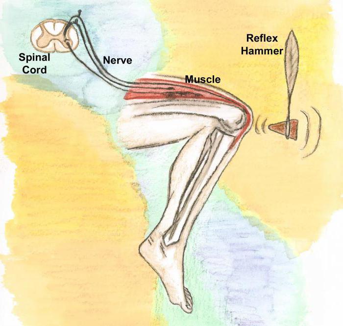
The entire doctrine of reflexes, from the studies of R. Descartes to the fundamental discoveries of I.P. Pavlov and I.M.Sechenov, is based on the notion of nervous activity as an adequate response of the body to the effects of stimuli of the external and internal environment, carried out with the participation of the central nervous systems of the brain and spinal cord. Whatever the answer, simple, for example, a knee-jerk reflex, or as super-complex as speech, memory or thinking, its first link is reception - the perception and discrimination of stimuli by their strength, amplitude, intensity.
Such differentiation is carried out by sensory systems, which IP Pavlov called "the tentacles of the brain." In each analyzer, the receptor performs the functions of antennas that capture and probe environmental stimuli: light or sound waves, chemical molecules, and physical factors. The physiologically normal activity of all sensory systems, without exception, depends on the work of the first department, called peripheral, or receptor. All reflex arcs (reflexes) originate from it.
Mediators
These are biologically active substances that transfer excitation from one neuron to another in special structures - synapses. They are secreted by the axon of the first neurocyte and, acting as an irritant, cause nerve impulses in the receptor endings of the next nerve cell. Therefore, the structure and functions of mediators and receptors are closely interrelated. Moreover, some neurocytes are capable of secreting two or more transmitters, for example, glutamic and aspartic acids, adrenaline and GABA.