This article will consider such structures of eukaryotic cells as chromosomes, the structure and functions of which are studied by a branch of biology called cytology.
Discovery story
Being the main components of the cell nucleus, chromosomes were discovered in the 19th century by several scientists. The Russian biologist I. D Chistyakov studied their mitosis (cell division) process , the German anatomist Valdeyer discovered them during the preparation of histological preparations and called them chromosomes, that is, stained bodies for the quick reaction of these structures when interacting with the fuchsin organic dye.
Fleming summarized the scientific facts about the function of the chromosomes in cells with a shaped nucleus.
The external structure of chromosomes
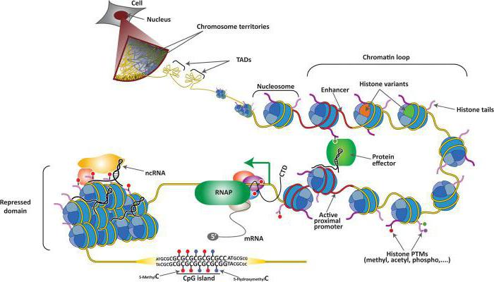
These microscopic formations are located in the nuclei - the most important organelles of the cell, and serve as a place of storage and transmission of hereditary information of this organism. Chromosomes contain a special substance - chromatin. It is a conglomerate of thin threads - fibrils and granules. From a chemical point of view, this is a combination of linear DNA molecules (about 40% of them) with specific histone proteins.
The complexes, which include 8 peptide molecules and DNA strands twisted on protein globules, like on coils, are called nucleosomes. The deoxyribonucleic acid site forms 1.75 revolutions around the core and has an ellipsoid shape of approximately 10 nanometers in length and 5-6 in width. The presence of these structures (chromosomes) in the nucleus serves as a systematic sign of the cells of eukaryotic organisms. It is in the form of nucleosomes that chromosomes perform the function of preserving and transmitting all genetic traits.
The dependence of the structure of the chromosome on the phase of the cell cycle
If a cell is in a state of interphase, which is characterized by its growth and intense metabolism, but the absence of division, then the chromosomes in the nucleus have the form of thin despiralized strands - chromonomes. Usually they are intertwined, and it is impossible to visually separate them into separate structures. At the time of the onset of cell division, which is called mitosis in somatic cells and meiosis in sex cells, the chromosomes begin to spiral and thicken, becoming clearly distinguishable under a microscope.
Chromosome Organization Levels
The units of heredity are chromosomes, a science of genetics studies in detail. Scientists have found that a nucleosome strand containing DNA and histone proteins form a first-order helix. The tight packing of chromatin occurs due to the formation of a higher order structure - the solenoid. It organizes itself and condenses into an even more complex supercoil. All of the above levels of chromosome organization pass during the preparation of the cell for division.
It is in the mitotic cycle that the structural units of heredity, consisting of genes containing DNA, are shortened and thickened by about 19 thousand times compared with the filamentous chromonemes of the interphase period. In such a compact form, the chromosomes of the nucleus, whose functions are to transmit the hereditary traits of the body, become ready for division of somatic or germ cells.
Chromosome morphology
The functions of chromosomes can be explained by studying their morphological features, which are best seen in the mitotic cycle. It is proved that even in the synthetic stage of interphase, the DNA mass in the cell doubles, since each of the daughter cells formed as a result of division must have the same amount of hereditary information as the original maternal one. This is achieved as a result of the reduplication process - DNA self-doubling, which occurs with the participation of the DNA polymerase enzyme.
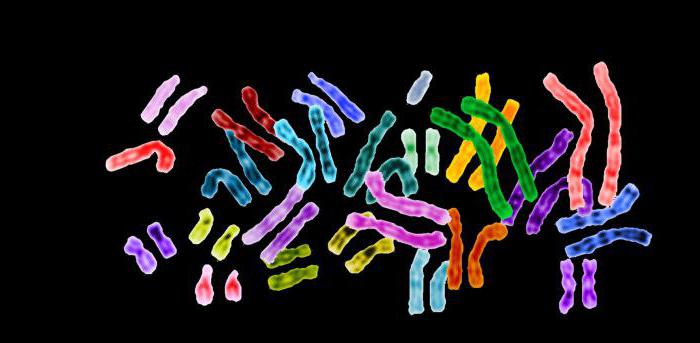
In cytological preparations prepared at the time of the metaphase of mitosis, in plant or animal cells under a microscope, it is clearly seen that each chromosome consists of two parts, called chromatids. In the subsequent phases of mitosis - anaphase and, especially, telophase - they are completely separated, as a result of which each chromatid becomes a separate chromosome. It contains a continuously condensed DNA molecule, as well as lipids, acidic proteins and RNA. Of the minerals, magnesium and calcium ions are present in it.
Auxiliary structural elements of the chromosome
In order for the chromosome functions in the cell to be fully realized, these units of heredity have a special device - the primary constriction (centromere), which never spirals. It is she who divides the chromosome into two parts, called the shoulders. Depending on the location of the centromere, geneticists classify the chromosomes into equal arms (metacentric), unequal shoulders (submetacentric) and acrocentric. On the primary constrictions, special formations are formed - kinetochores, which are disk-shaped protein globules located on both sides of the centromere. The kinetochores themselves consist of two sections: the outer ones are in contact with microfilaments (threads of the fission spindle), attaching to them.
Due to the reduction of filaments (microfilaments), a strictly ordered distribution of the chromatids that make up the chromosome between daughter cells is carried out. Some chromosomes have one or more secondary constrictions that do not participate in mitosis, since the threads of the fission spindle cannot join them, but it is these areas (secondary constrictions) that provide control over the synthesis of nucleoli - organelles, which are responsible for the formation of ribosomes.
What is a karyotype
Well-known genetic scientists Morgan, N. Koltsov, Setton at the beginning of the 20th century scrupulously studied chromosomes, their structure and functions in somatic and germ cells - gametes. They found that each cell of all biological species is characterized by a certain number of chromosomes having a specific shape and size. It was proposed that the entire set of chromosomes in the nucleus of a somatic cell be called a karyotype.
In popular literature, a karyotype is often identified with a chromosome set. In fact, these are not identical concepts. For example, in humans, the karyotype is 46 chromosomes in the nuclei of somatic cells and is indicated by the general formula 2n. But cells such as hepatocytes (liver cells) have several nuclei, their chromosome set is designated as 2n * 2 = 4n or 2n * 4 = 8n. That is, the number of chromosomes in such cells will be more than 46, although the hepatocyte karyotype is 2n, i.e. 46 chromosomes.
The number of chromosomes in germ cells is always two times less than in somatic cells (in body cells), such a set is called haploid and is designated as n. All other cells in the body have a 2n kit called diploid.
Morgan's chromosome theory of heredity
American geneticist Morgan discovered the law of linked inheritance of genes, conducting experiments on the hybridization of fruit flies-Drosophila. Thanks to his studies, the function of germ cells chromosomes was studied. Morgan proved that genes located at neighboring loci of the same chromosome are inherited primarily together, that is, linked. If the genes are located on the chromosome far from each other, then crossover is possible between sister chromosomes - the exchange of sites.
Thanks to Morgan's research, genetic maps have been created with which they study the functions of chromosomes and widely use them in genetic counseling to solve questions about possible pathologies of chromosomes or genes that lead to hereditary diseases in humans. The importance of the conclusions made by the scientist is difficult to overestimate.
In this article, we examined the structure and functions of the chromosomes that they perform in the cell.