The heart in the human body is a vital organ. Its work can be compared to a pump. Thanks to the heart, blood is pumped into the arteries and continuously moves through the vessels. The named organ functions throughout the life of a person. Over 70 years, it performs approximately 2-3 billion contractions and pumps over 170 million liters of blood. So how does the heart work? What are its functions?
The location and size of the heart
The main organ of the human body is located in the center of the chest. Most of the heart is located in the left half of the body, and the smaller - in the right. The organ lies in the pericardial sac. It is also called the pericardium. This is a dense bag that blocks the heart from other internal organs and does not allow it to move and overstretch at the time of physical exertion.
The size of the heart is quite small. Each person has about a fist. However, the size and weight of the organ may vary. Parameters increase with some ailments. The size and weight of the heart also increase in those individuals who for a long period of time are involved in sports or intense physical labor.
Organ structure
Let's see how the heart works. The walls of this organ form three layers:
- Epicardium. This is a thin membrane outer layer of the heart wall.
- Myocardium. Under this term, specialists understand the middle layer, which is responsible for muscle contractions of the heart.
- Endocardium. This is a membrane that limits the internal system of the heart.
This vital organ consists of two parts separated by a partition - a thick muscle wall. Each half includes two cameras. The upper sections (right and left) are called the atria, and the lower - the ventricles. Each chamber plays a special role in the blood circulation.
Atria
Considering how the heart is arranged, it is worth talking about the atria - the thin-walled chambers of the heart. They are located above the ventricles and are separated from them by atrioventricular valves. Allocate the right and left atria. The upper right chamber of the organ is the place where the hollow veins and veins of the heart enter. Based on this information, it can be concluded that this atrium receives venous blood, devoid of oxygen.
The upper left organ chamber is smaller in size than the right one. Four openings of pulmonary veins open in it. Of these, fresh blood, saturated with oxygen and ready for further distribution throughout the human body, enters the left atrium.
Ventricles
In the picture, which shows how a person’s heart is arranged (photo below), you can see the right and left ventricles. They form the main muscle mass of the organ. It should be noted that the left camera is more massive and powerful compared to the right one. Venous blood enters the right ventricle from the right atrium. When the heart muscle contracts, it goes to the lungs through the pulmonary valve. The tricuspid valve, also called the tricuspid valve, prevents the return of blood to the upper chamber.
The left ventricle receives oxygenated blood from the left atrium. It enters through the mitral (bicuspid) valve. With contractions of the muscles of the lower left chamber, blood is pushed into the aorta through the aortic valve. Further, it is carried throughout the human body.
Heart work
When considering how the heart is arranged, it is necessary to study the work of the organ. The ventricles and atria can be either in a relaxed (diastolic) or shortened (systolic) state. Relaxations and contractions of the heart occur in a certain sequence:
- Systole of the atria. The contraction of the upper chambers of the organ is the beginning of the cardiac cycle. This phase lasts 0.1 s. During systole, the flap valves open. All blood from the atria is sent to the ventricles. After the reduction of the upper chambers, the relaxation phase begins.
- Ventricular systole. The contraction of the lower parts of the heart lasts 0.3 s. Lunate (pulmonary and aortic) and leaf valves at the beginning of the phase are closed. The muscles of the ventricles contract. Because of this, the pressure in the cavities rises. As a result, the blood goes to the atria. There the pressure is lower. However, leaflets prevent the flow of blood in this direction. Their folds cannot turn inside the atria. At this moment, the lunar valves open . Blood begins to move along the pulmonary artery and aorta.
- Diastole. The ventricles relax after contraction. This phase lasts 0.4 s. During the resting period of the organ, blood flows from the veins into the atria and partially penetrates into the ventricles. When a new cycle begins, the remnants of blood from the upper chambers of the organ are pushed into its lower sections.
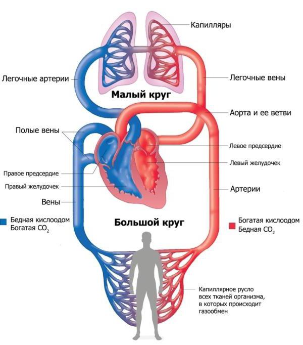
Considering how the heart is arranged and how it works, it is worth talking about the circles of blood circulation - large and small. The first of these begins with the aorta. Oxygen-enriched blood enters into it from the left ventricle. From the largest arterial vessel, it flows through arteries, arterioles, capillaries, delivering oxygen to all cells and freeing them from accumulated carbon dioxide. As a result, the venous blood leaves the capillary network. First, it moves through the venules, and then through the veins and vena cava. As a result, she falls into the right atrium, and from it is sent to the right ventricle.
The pulmonary circulation begins with a pulmonary artery emerging from the lower right chamber of the heart. Venous blood enters the lungs, moves through the arteries, arterioles, and the thinnest capillaries located in these organs. As a result, she gets to the alveoli - tiny bubbles that are filled with air. Blood absorbs oxygen, is purified from carbon dioxide and enters the veins. These blood vessels go to the left atrium. From it, blood is pushed into the left ventricle. Further, everything is repeated again. Blood begins to move in a large circle of blood circulation.
Body functions
Having examined how the heart works, we can name its functions. One of them is reservoir. The vital organ of the human body during the period of relaxation of the heart muscle serves as a cavity for the accumulation of the next portion of blood coming from the blood vessels into the atria. The second function of the heart is injection. It consists in the release of blood into the small and large circles of blood circulation with the contraction of the ventricles.
How a person’s heart works, everyone should know. Everyone needs to have information about how his body is structured, what processes are taking place in it. The well-being and human health depend on the work of the heart. Due to the functioning of this organ, blood is carried throughout the body, supplies all organs and tissues with oxygen, biologically active substances, energy and takes carbon dioxide and excretion products from them.