Cataracts are a pathological condition in which a clouding of the lens of the eye occurs, leading to a progressive decrease in vision. When a certain age is reached, some changes occur in the human lens, which are reflected in the compaction of the nucleus and its delimitation from the cortical zone. The immature cataract in ICD-1010 is encrypted with code H 26.
The first signs and diagnosis of cataracts
The separation of the cataract maturation process into 4 stages, as well as into 2 main forms: cortical and nuclear. Cortical cataract is manifested by clouding, starting from the periphery and going to the center, accompanied by a progressive decrease in visual acuity.
The main complaints, in addition to a drop in visual acuity, will be:
- a feeling of veil before the eyes;
- blurred gaze;
- frequent change of glasses without much effect.
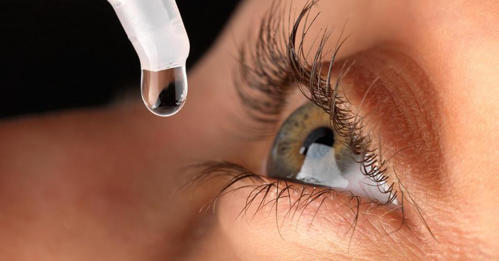
Diagnosis of cataracts is carried out by biomicroscopy (that is, examination of the eye on a slit lamp). The doctor can determine exactly which cataract is present, as well as determine its stage. In its development, cataract passes through several stages:
- Initial cataract is characterized by the appearance of flat opacities. Turbidity located on the periphery of the lens is more intense. When clouding is in the cortex, initial cataract may not reduce visual acuity.
- Immature age-related cataracts - opacities are moving in all directions, capturing an increasingly large surface of the lens and becoming more intense. Visual acuity in this case may decrease to tenths and hundredths.
- Mature cataract is detected when the entire cortex is already becoming cloudy. Vision with such clouding may decrease up to light projection (as a rule, correct light projection, in view of the retina’s preserved function).
- Overripe cataracts are a degeneration and decomposition of the lens fibers. There is a liquefaction of the substance of the lens, in connection with which the folding of the capsules appears. The color of the bark changes to milky. The nucleus, being a dense and heavy formation, can go down and only its upper edge will be noticeable during biomicroscopy.
Conservative treatment
Conservative treatment is a controversial issue in ophthalmology, some authors note an objective slowdown in cataract progression, while others, on the contrary, say that there is no effect from the drugs used.
Nevertheless, the following groups of drugs for the treatment of cataracts are present in the pharmaceutical market:
- Drops containing salts of K, Mg, Ca, Li, J and others, necessary for normal water and electrolyte metabolism.
- Means that carry out the correction of the lens metabolism, which include biological products, hormones and vitamin complexes.
- Preparations that include organic compounds that contribute to the normalization of metabolic reactions.
- Vitamins: riboflavin, glutamic acid, ascorbic acid, cysteine, taufon or taurine.
Surgery
Only surgical treatment of cataracts can cure the patient. It consists in the removal (extraction) of the clouded lens with further installation of an artificial lens in its place. There are several types of cataract extraction:
- The removal of the lens is done together with the capsule (IEK). Removing part of the anterior capsule of the lens, then there is a fragmentation of the nucleus, its aspiration, after which all other masses are aspirated. The back capsule is not damaged. (EEC)
- Removal of the lens through a small incision through the use of an ultrasonic emitter (ultrasound FEK).
- The destruction of the nucleus and cortex using laser energy and their removal using vacuum (LEK).
Aphakia Correction
After the operation of an immature cataract, namely the removal of the lens, the patient’s vision also remains low, due to the lack of a natural lens of 19 diopters. Such an eye is called aphakic and has certain signs:
- deep front camera;
- iris tremor - iridodenesis;
- hyperopic refraction.
A similar situation can be solved in several ways:
- spectacle correction (collecting lenses);
- contact correction (soft contact lenses);
- correction with an intraocular lens.
An intraocular lens (IOL) is an artificial collecting lens made from inert materials and placed inside the eyeball to correct aphakia. The main parts of the IOL are optics and haptics.
Complications of Surgical Treatment
In most cases, at the present stage of development of microsurgical technology, operations on the lens are safe, however, complications, although not often, still occur. The vast majority of operations are performed for cataracts using the ultrasonic method of nuclear destruction. The ultrasonic wave emanating from the emitter is capable of damaging the surrounding tissue. In order to avoid this, manipulations inside the anterior chamber of the eye are performed with the introduction of viscoelastic. It is a liquid with a very high viscosity. A similar feature allows it to well absorb the waves emanating from the emitter.
The most common non-specific complication of immature cataract is a postoperative inflammatory reaction. Any operation is accompanied by an inflammatory reaction of varying severity, as a natural tissue response to damage.
Possible consequences
Clinical manifestations of postoperative inflammation include:
- artifactic cell precipitates;
- clouding of the posterior lens capsule;
- transient postoperative hypertension, glaucoma;
- Hyphema, cystic macular edema;
- sluggish fibro-plastic uveitis;
- the formation of posterior synechiae; pupillary membrane.
Specific complications of such an operation with immature cataracts will be problems at any stage of its conduct. There may be complications at the stage of cutting the cornea, when separating the nucleus from the entire lens, when creating a window in the anterior capsule of the lens, leaving the nucleus in the anterior or posterior chamber of the eye, and problems in setting up the IOL.