Of great importance for the perception of the world are the organs of vision. Thanks to the eyes, people and animals receive 90% of the information. Therefore, problems with the organ of vision are always an occasion to seek help from a specialist. Only through the necessary examinations can we understand why the violation occurred. Diagnosis of ocular pathologies includes measuring visual acuity, ophthalmoscopy, examination of retinal vessels, and computer perimetry. Each of these studies is important for identifying diseases. Thanks to this method, you can find out exactly which site fell out of vigorous activity.
Description of computer perimetry
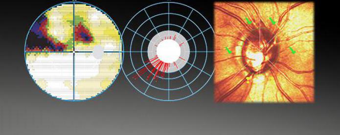
Computer perimetry is a research method due to which a change in the field of view can be detected. Normally, a person sees not only what is directly in front of him, but also another part of the surrounding objects located on the sides. This function is carried out thanks to peripheral vision, for which the brain is responsible. With various ophthalmic and neurological pathologies, loss of visual fields occurs . Such violations include hemianopsia. The loss of one or more visual fields and its replacement with a white veil is called scotoma. Computer perimetry of the eye allows you to assess the number and size of defects. Also, thanks to it, it is possible to diagnose those visual disturbances that are at an early stage and are not yet clinically manifested. Previously, there were other devices for cattle detection. Nevertheless, computer perimetry differs from them in a higher accuracy in calculating the boundaries of the field of view and existing defects. This diagnostic method is a safe and non-invasive procedure.
What is the study of visual fields for?
The narrowing or total disappearance of the visual field is a serious violation. The same applies to the loss of its plots - cattle. In some cases, the pathology is not considered ophthalmic, but refers to diseases of the brain. Therefore, we can distinguish the following indications for computer perimetry:
- Dystrophy of the retina.
- Acid or alkali damage to the organ of vision, thermal burns.
- Retinal hemorrhage.
- Tumor lesions of the organ of vision.
- Increases in intraocular pressure - glaucoma.
- Retinal detachment.
- Inflammation or damage to the optic nerve.
- Brain injuries.
- Hemorrhagic and ischemic stroke.
- Retinopathies caused by arterial hypertension and diabetes.
All these conditions are quite dangerous, since in advanced cases they can lead to complete blindness.
Computer Perimetry Technique
To explore the field of view, it is necessary to fix the gaze on a certain subject. Everything that a person “catches” with his eye outside this image is carried out with the help of peripheral vision. It is worth remembering that the study of vision is contraindicated in some situations. Among them:
- State of alcohol or drug intoxication.
- Emotional lability.
- Lag in mental development.
In all these conditions, the patient is not able to clearly concentrate his gaze and follow the instructions of an ophthalmologist. Computer perimetry is based on the study of the capabilities of the organ of vision in the formulation of a number of tasks. The patient is seated for a special device having an optical system. Each eye is checked separately, while the second is covered with a shutter. First of all, the patient fixes his gaze on one subject. In this way, the breadth of the visual fields is estimated. After that, other objects appear around the main image - objects that are different in light and brightness. In this case, the gaze should also be fixed. Further, the images on the periphery move in space. Thanks to this method, it is possible to evaluate not only the size of the fields of view, but also the susceptibility to colors, light, movement.
Types of computer perimetry of the eye
Depending on what kind of "picture" is depicted on the periphery, several types of research are distinguished. In most cases, they are all applied in turn. This helps to identify a large number of deviations from the norm and get an idea of visual function. Types of computer perimetry:
- Static The patient fixes his gaze on the white dot located in the center of the device, and the visual fields at this moment are projected on a rounded surface. To accurately record readings, lighting is constantly changing.
- Kinetic. The patient needs to monitor an object that is in motion. While the subject is approaching and moving away from the eyes, the device captures the necessary indicators.
- Campimetry. The examinee must observe a moving white dot located inside the dark square. The device evaluates the boundaries at which the object disappears and reappears.
- Amsler test. The patient is asked to focus his eyes on the middle of the picture (grille). If the person being examined sees straight lines, then there are no problems with the retina.
Computer perimetry: decoding of this method
After the study, the results are recorded on a map used by ophthalmologists. Normally, the lower and the inner border should be 60, the top - 50, and the outer - 90 degrees. The presence of physiological cattle is not considered a pathology, as they arise due to a blind spot located on the retina. If the loss of fields is large or multiple, it is associated with diseases of the organ of vision or the brain. Hemianopsia indicates pathology of the optic nerve. By the number and nature of livestock, one can judge diseases such as migraine and glaucoma.
What ophthalmological clinics in St. Petersburg carry out the study?
In any large regional center, you can be examined for the presence of pathology of vision. The northern capital is no exception. Where can I get computer perimetry in St. Petersburg? The following ophthalmological clinics are known (in St. Petersburg), having an apparatus for this study:
- Oncoscreening Center.
- The world of health.
- Clinic Medem.
- Alpha Medic.
- "Family doctor".
- Research Institute of Experimental Medicine.
The cost of computer perimetry is from 400 to 1200 rubles.