To interact with the outside world, a person needs to receive and analyze information from the external environment. For this, nature has endowed it with sensory organs. There are six of them: eyes, ears, tongue, nose, skin and vestibular apparatus. Thus, a person forms an idea about everything that surrounds him and about himself as a result of visual, auditory, olfactory, tactile, taste and kinesthetic sensations.
It can hardly be argued that some sense organ is more significant than the rest. They complement each other, creating a complete picture of the world. But the fact that most of all information is up to 90%! - people perceive through the eyes - this is a fact. In order to understand how this information enters the brain and how its analysis occurs, you need to imagine the structure and functions of the visual analyzer.
Features of the visual analyzer
Thanks to visual perception, we learn about the size, shape, color, relative position of the objects of the world, their movement or immobility. This is a complex and multi-step process. The structure and functions of the visual analyzer - a system that receives and processes visual information, and thereby provides vision - are very complex. Initially, it can distinguish peripheral (perceiving the initial data), conducting and analyzing parts. Information is obtained through the receptor apparatus, which includes the eyeball and auxiliary systems, and then it is sent using the optic nerves to the corresponding centers of the brain, where it is processed and visual images are formed. All departments of the visual analyzer will be considered in the article.
How the eye works. The outer layer of the eyeball
The eyes are a paired organ. Each eyeball resembles a slightly flattened ball in shape and consists of several shells: the outer, middle and inner, surrounding the fluid-filled eye cavities.
The outer shell is a dense fibrous capsule that preserves the shape of the eye and protects its internal structures. In addition, six motor muscles of the eyeball are attached to it. The outer shell consists of a transparent front part - the cornea, and the back, opaque - sclera.
The cornea is a refractive medium of the eye, it is convex, has the appearance of a lens and consists, in turn, of several layers. There are no blood vessels in it, but there are many nerve endings. A white or bluish sclera, the visible part of which is usually called the white of the eye, is formed from connective tissue. Muscles are attached to it, providing turns of the eyes.
The middle layer of the eyeball
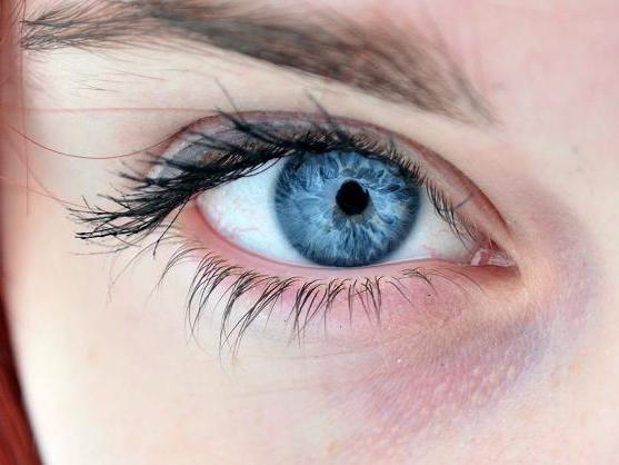
The middle choroid is involved in metabolic processes, providing nutrition to the eye and the withdrawal of metabolic products. The front, most noticeable part of it is the iris. The pigment substance located in the iris, or rather, its amount, determines the individual shade of a person’s eyes: from blue, if it is small, to brown, if enough. If there is no pigment, as happens with albinism, then the plexus of the vessels becomes visible, and the iris becomes red.

The iris is located immediately behind the cornea, its basis is muscles. The pupil - a rounded hole in the center of the iris - thanks to these muscles, it regulates the penetration of light into the eye, expanding in low light and narrowing in too bright. The continuation of the iris is the ciliary (ciliary) body. The function of this part of the visual analyzer is to produce fluid that feeds those parts of the eye that do not have their own vessels. In addition, the ciliary body directly affects the thickness of the lens through special ligaments.
In the posterior part of the eye in the middle layer is the choroid, or the choroid itself, which is almost entirely composed of blood vessels of different diameters.
Retina
The inner, thinnest layer is the retina, or the retina, formed by nerve cells. Here is the direct perception and primary analysis of visual information. The back of the retina consists of special photoreceptors called cones (there are 7 million) and rods (130 million). They are responsible for the perception of objects by the eye.
Cones are responsible for color recognition and provide central vision, allow you to see the smallest details. The sticks, being more sensitive, enable a person to see in black and white colors in low light conditions, and are also responsible for peripheral vision. Most cones are concentrated in the so-called yellow spot opposite the pupil, slightly above the entrance of the optic nerve. This place corresponds to maximum visual acuity. The retina, as well as all departments of the visual analyzer, by the way, has a complicated structure - 10 layers are distinguished in its structure.
The structure of the eye cavity
The eye nucleus consists of the lens, vitreous body and chambers filled with fluid. The lens looks like a transparent lens convex on both sides. It has neither vessels, nor nerve endings and is suspended from the processes of the surrounding ciliary body, the muscles of which change its curvature. This ability is called accommodation and helps the eye focus on near or, conversely, distant objects.
Behind the lens, adjacent to it and further to the entire surface of the retina, is the vitreous. This is a transparent gelatinous substance that fills most of the volume of the organ of vision. 98% of this gel-like mass is water. The purpose of this substance is to conduct light rays, compensate for intraocular pressure drops, maintain the constancy of the shape of the eyeball.
The anterior chamber of the eye is bounded by the cornea and iris. It connects through the pupil to a narrower posterior chamber, extending from the iris to the lens. Both cavities are filled with intraocular fluid, which freely circulates between them.
Refraction of light
The system of the visual analyzer is such that initially the rays of light are refracted and focused on the cornea and pass through the anterior chamber to the iris. Through the pupil, the central part of the light flux hits the lens, where it focuses more accurately, and then through the vitreous to the retina. The image of the object is projected on the retina in a reduced and, moreover, inverted form, and the energy of light rays by photoreceptors is converted into nerve impulses. Information is then passed through the optic nerve to the brain. The place on the retina through which the optic nerve passes is devoid of photoreceptors, therefore it is called a blind spot.
The motor apparatus of the organ of vision
The eye, in order to respond to stimuli in a timely manner, must be mobile. Three pairs of oculomotor muscles are responsible for the movement of the visual apparatus: two pairs of straight and one oblique. These muscles are perhaps the fastest in the human body. The oculomotor nerve controls the movement of the eyeball. It connects four of the six eye muscles to the nervous system , ensuring their proper functioning and coordinated eye movements. If the oculomotor nerve for some reason ceases to function normally, this is expressed in various symptoms: strabismus, drooping eyelids, double vision, dilated pupil, accommodation disturbances, bulging eyes.
Eye protective systems
Continuing such a voluminous topic as the structure and functions of the visual analyzer, it is impossible not to mention those systems that protect it. The eyeball is located in the bone cavity - the orbit, on a shock-absorbing fat pad, where it is reliably protected from shock.
In addition to the orbit, the protective apparatus of the organ of vision includes the upper and lower eyelids with eyelashes. They protect the eyes from falling from the outside of various objects. In addition, the eyelids help evenly distribute tear fluid on the surface of the eye, remove tiny particles of dust when blinking from the cornea. Eyebrows also to some extent perform protective functions, protecting the eyes from sweat draining from the forehead.
Lacrimal glands are located in the upper outer corner of the orbit. Their secret protects, nourishes and moisturizes the cornea, and also has a disinfecting effect. Excess fluid flows through the tear duct into the nasal cavity.
Further conducting and final processing of information
The conductor section of the analyzer consists of a pair of optic nerves that exit from the orbits and enter special channels in the cranial cavity, further forming an incomplete cross, or chiasm. Images from the temporal (outer) part of the retina remain on the same side, and from the inner, nasal - they intersect and are transmitted to the opposite side of the brain. As a result, it turns out that the right visual fields are processed by the left hemisphere, and the left - by the right. Such an intersection is necessary for the formation of a three-dimensional visual image.
After the intersection, the nerves of the conductor section continue in the optic tracts. Visual information enters the part of the cerebral cortex that is responsible for its processing. Such a zone is located in the occipital region. There, the final transformation of the received information into a visual sensation takes place. This is the central part of the visual analyzer.
So, the structure and functions of the visual analyzer are such that disturbances in any of its sections, whether it be perceiving, conducting or analyzing zones, entail a failure of its operation as a whole. This is a very multifaceted, subtle and perfect system.
Violations of the visual analyzer - congenital or acquired - in turn, lead to significant difficulties in understanding reality and limiting opportunities.