Evoked visual potentials are biological potentials that appear in the cerebral cortex as a reaction to exposure to light on the retina.
A bit of history
They were first described by E.D. Adrian in 1941, but they were steadily fixed after Davis and Galambos advanced a methodology for summing potentials in 1943. Then, the VEP registration method was widely used in a clinic where the functional position of the visual pathway was studied in patients in the ophthalmoneurological sphere. To register VIZ, they use specialized standard electrophysiological systems based on the work of modern computers.
A metal plate, that is, an active electrode, is placed on the patient’s head two centimeters above the occipital protuberance along the midline above the area where the visual striatal cortex is projected onto the cranial arch. An indifferent second electrode is mounted on the earlobe or mastoid process. A ground electrode is attached to the earlobe of the other ear or to the skin in the middle of the forehead. How is vision testing performed on a computer? As a stimulant, either a flash of light (flare VIZ) or reversible patterns from a monitor (pattern-VIZ) are used. The stimulating field of view is approximately fifteen degrees in size. Studies are carried out without enlarged pupils. The age of the person undergoing the procedure also plays a role. We will understand how a person sees.
More about the concept
VEP are the bioelectric response of the visual regions located on the cerebral cortex, and the thalamocortical pathways and subcortical nuclei. Wave generation of VEP is also associated with generalized mechanisms of brain activity of a spontaneous nature, which is recorded on the EEG. Responding to the effect of light on the eyes, PTA show bioelectric activity of the mainly macular sphere of the retina, which is due to its large representation in the visual cortical centers in comparison with the parts of the retina located on the periphery.
How is registration done?
Evoked visual potentials are recorded in the form of oscillations of an electric potential of a sequential nature or components that differ in polarity: the negative potential, or N, has an upward direction, a positive potential, that is, P, downward. The characteristic of VIZ contains a form and two quantitative indicators. The potential VIZ in size is normally much smaller (up to about 40 μV) in comparison with the waves of the electroencephalogram (reaches 100 μV). Latency is determined using the time period from the moment the light stimulus is turned on until the maximum potential indicator of the cerebral cortex is reached. Most often, the potential reaches its maximum value after 100 ms. If there are various kinds of pathology of the visual pathway, then the form of the VEP changes, the amplitude of the components decreases, the latency lengthens, that is, the time increases during which the impulse passes to the cerebral cortex along the visual pathway.
In what proportion is the visual zone? It is located in the occipital lobe of the brain.
Varieties
The nature of the components in the VIZ and their sequence is quite stable, but at the same time, the temporal characteristics and amplitude normally vary. This is determined by the conditions in which the study is conducted, the specifics of the light stimulus, and the application of electrodes. During stimulation of the half-fields of vision and the reverse frequency from one to four times per second, a phase transient VEP is recorded, in which three components are sequentially distinguished - N 70, P 100 and N 150. The frequency of reversal with an increase of more than four times per second causes the appearance of a rhythmic the total response in the cerebral cortex in the form of a sinusoid, which is called the VIZ steady state state. These potentials differ from phase potentials in that they lack serial components. They look like a rhythmic curve, with alternating drops and rises in potential.
Normal indicators of evoked visual potentials
VEP analysis is carried out according to the amplitude of potentials, measured in microvolts, according to the form of recording and the time period from exposure to light to the occurrence of peak waves of the CEM (calculation in milliseconds). Also pay attention to the difference in the amplitude of the potential and the latency during light stimulation in the right and left eye in turn.
In VIZ (which is interesting in ophthalmology for many) of the phase type when reversing with a low frequency of a chess pattern or in response to a light flash with special constancy, P 100, a positive component, is released. The duration of the latent period of this component varies normally from ninety-five to one hundred and twenty milliseconds (cortical time). The preceding component, that is, N 70, is from sixty to eighty milliseconds, and N 150 is from one hundred fifty to two hundred. Late P 200 is not recorded in all cases. This is how an eye test on a computer passes.
Since the amplitude of the VIZ is distinguished by its variability, when taking into account the results of the study, it has a relative value. Normally, its values with respect to P 100 fluctuate in an adult from fifteen to twenty-five μV, higher potential values in children - up to forty μV. On the stimulation pattern, the amplitude of the VIZ is slightly lower and is determined by the magnitude of the pattern. If the value of the squares is greater, then the potential is higher, and vice versa.
Thus, the evoked visual potentials are a reflection of the functional state of the visual pathways and allow obtaining quantitative information during the course of the study. The results make it possible to diagnose pathologies of the visual pathway in patients of the neuroophthalmological region.
This is how a person sees.
Topographic mapping of biopotentials of the brain of the head according to VIZ
Topographic mapping of the brain brain biopotentials using VES multichannel records biopotentials from various areas of the brain: parietal, frontal, temporal and occipital. The results of the study are transmitted to the monitor screen as topographic maps in a color that varies from red to blue. Thanks to topographic mapping, the amplitude value of the VIZ potential in ophthalmology is shown. What is it, we explained.
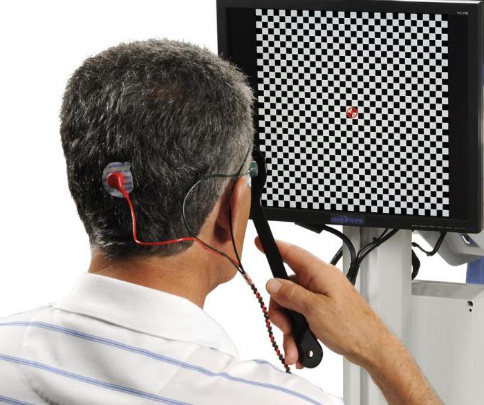
A special helmet with sixteen electrodes is put on the patient’s head (the same as for EEG). Electrodes are placed on the scalp at specific points in the projection: parietal, frontal over the left and right hemispheres, temporal and occipital. Processing and registration of biopotentials is carried out using specialized electrophysiological systems, for example, a Neurocartograph from MBN. Using this technique, it becomes possible to conduct an electrophysiological differential diagnosis in patients. In acute retrobulbar neuritis, on the contrary, bioelectrical activity is noted, which is expressed in the nape of the head, and there is almost complete absence of excited areas in the frontal lobe of the brain.
Diagnostic value of evoked visual potentials in various pathologies
In physiological and clinical studies, if the visual acuity is high enough, it is best to use the method of registering physical VEP for reversion.
In clinical and physiological studies with a sufficiently high visual acuity, it is preferable to use the method of registering physical VEP for reversible chess patterns. These potentials are quite stable in terms of amplitude and temporal properties, are well reproducible and are sensitive to various pathologies in the visual pathways.
On an outbreak, VEPs are more varied and less sensitive to changes. This method is used for a serious decrease in visual acuity in a patient, lack of gaze fixation, with impressive opacification of ophthalmic means, pronounced nystagmus and in young children.
The following criteria are involved in the vision test:
- lack of response or a large decrease in amplitude;
- longer latency of all climaxes of potentials.
When recording evoked visual potentials, it is necessary to take into account the norm by age, especially with regard to the study of children. Interpreting the information on the registration of VEP in early childhood with pathologies of the visual pathways, one should take into account the characteristic features of the electrocortical reaction.
Two phases in the development of VEP are distinguished, which are recorded in response to pattern reversal:
- fast - from birth to six months;
- slow - from six months to the puberty.
In the first days of life in children, VEP is recorded.
Topical diagnosis of brain pathologies
What does EEG show? At the chiasmal level, the pathology of the visual pathways (tumors, injuries, optochiasmal arachnoiditis, demyelinating processes, aneurysms) decreases in the amplitude of potentials, latency increases, some elements of the VIZ drop out. There is an increase in changes in VIZ simultaneously with the progression of the lesion. The prechiasmal region of the optic nerve is involved in the pathological process, which is confirmed ophthalmoscopically.
Retrochiasmal pathologies are distinguished by interhemispheric asymmetry of visual potentials and are better traced with the multi-channel type of recording, topographic mapping.
Chiasmal lesions are characterized by an asymmetry of VIZ of a crossed nature, expressed in significant changes in biopotentials in the brain on the opposite side of the eye, which has reduced visual functions.
During the analysis of VEP, it is necessary to take into account the hemianopic loss of the visual field. In this regard, with chiasmal pathologies, light stimulation of half of the visual field increases the sensitivity of the method, which allows to identify the distinguishing signs between dysfunction in the fibers of vision, which come from the nasal and temporal parts of both eye retinas.
At the retrochiasmal level of visual pathway defects (Graziole beam, optic tract, visual region of the cerebral cortex), a one-sided dysfunction is observed, manifested in the form of non-crossed asymmetry, which is expressed in pathological VEP, which have the same indicators when stimulating each eye.
The reason that the bioelectric activity of neurons in the central areas of the optic pathways decreases is homogeneous visual field defects. If they capture the macular region, then during the stimulation, half the field changes and gets the shape that is characteristic of central cattle. If primary visual centers are preserved, then VIZ can have normal values. What else shows the EEG?
Pathology of the optic nerve
If there are pathological processes in the optic nerve, then the most characteristic manifestation of them is an increase in latency of the main component of VEP P 100.
Optic neuritis from the affected eye along with an increase in latency is characterized by a decrease in the amplitude of the potentials and a change in components. That is, central vision is impaired.
Often, a W-shaped component of P 100 is registered, associated with a decrease in the functioning of the axial bundle of nerve fibers in the optic nerve. The disease progresses along with an increase in latency time of thirty to thirty-five percent, a decrease in amplitude, and formal changes in the components of the VEP. If the inflammatory process subsides in the optic nerve, and the visual functions increase, then the form of the VEP and the amplitude indicators normalize. The temporal characteristics of the VIZ remain increased for two to three years.
Optic neuritis, which develops against the background of multiple sclerosis, is determined even before the clinical symptoms of the disease are identified by the changes occurring in the VEP, which indicates the early involvement of the visual pathways in the pathological process.
The defeat of the optic nerve of a one-sided nature in this case has very significant differences in the latency of the component P 100 (twenty-one milliseconds).
Anterior and posterior ischemia of the optic nerve due to an acute defect in arterial circulation in those vessels that feed it is accompanied by a noticeable decrease in the amplitude of VEP from the side of the patient’s eye and not too high (by three milliseconds) increase in P 100 latency. In this case, the VEC of the healthy eye usually remain normal.
The stagnant disk at the initial stage is characterized by a decrease in the amplitude of visual evoked potentials (VEP) of a moderate nature and a slight increase in latency. If the disease progresses, then the disorders receive an even more tangible expression, which fully corresponds to the ophthalmoscopic picture.
In case of atrophy of the optic nerve of the secondary type after the transfer of ischemia, neuritis, stagnant disk and other pathological processes, a decrease in VEP amplitude and an increase in latency P 100 are also observed. Such changes can be characterized by different degrees of expression and appear independently of each other.
Pathological processes in the retina and choroid (serous central choriopathy, numerous forms of maculopathy, macular degeneration) contribute to an increase in the latency period and a decrease in the amplitude of potentials.
Often there is no correlation between a decrease in amplitude and an increase in the latency length of potentials.
Conclusion
So, we can conclude that although the method of analyzing VEP is not specific for determining any pathological process of the visual pathway, it is used for early diagnosis in the clinic of various kinds of eye diseases and to clarify the degree and level of damage. Of particular importance is the test for eye examination and in ophthalmic surgery.