The eyes are an important organ for the normal functioning of the body and a full life. The main function is the perception of light stimuli, due to which the picture appears.
Structural features
This peripheral organ of vision is located in a special cavity of the skull called the orbit. From the sides, the eye is surrounded by muscles, with the help of which it is held and moves. The eye consists of several parts:
- Directly the eyeball, which has the shape of a ball about 24 mm in size. It consists of a vitreous, crystalline lens and aqueous humor. All this is surrounded by three membranes: protein, vascular and reticular, located in the reverse order. Elements due to which the picture is obtained are located on the mesh shell. These elements are receptors that are sensitive to light;
- The protective apparatus, which consists of the upper and lower eyelids, orbits;
- Adnexa. The main components are the lacrimal gland and its ducts;
- The oculomotor apparatus, which is responsible for the movement of the eyeball and consists of muscles;
- Optic nerve.
Main functions
The main function that vision performs is to distinguish between various physical characteristics of objects, such as brightness, color, shape, size. In combination with the action of other analyzers (hearing, smell and others), it allows you to adjust the position of the body in space, as well as determine the distance to the object. That is why the prevention of eye diseases should be carried out with enviable regularity.
The presence of the pupil reflex
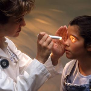
With the normal functioning of the organs of vision, with certain external reactions, the so-called pupillary reflexes occur, in which the pupil narrows or expands. The pupillary reflex, the reflex arc of which is an anatomical substrate of the pupil’s reaction to light, indicates the health of the eyes and the whole organism. That is why, for some diseases, the doctor first checks the presence of this reflex.
What is a reaction?
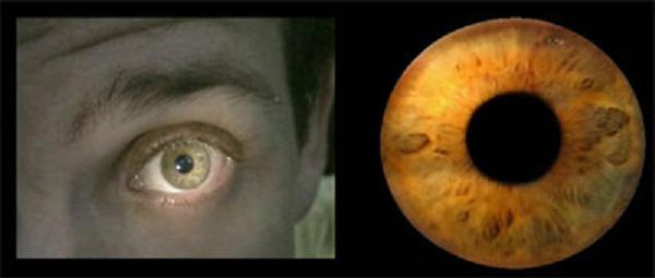
The reaction of the pupil or the so-called pupillary reflex (other names - the iris reflex, iritic reflex) is some change in the linear dimensions of the pupil of the eye. Narrowing, as a rule, is caused by contraction of the muscles of the iris, and the reverse process - relaxation - leads to the expansion of the pupil.
Possible reasons
This reflex is caused by a combination of certain stimuli, the main of which is a change in the level of illumination of the surrounding space. In addition, pupil resizing can occur for the following reasons:
- the effect of a number of medicines. That is why they are used as a way to diagnose the condition of drug overdose or excessive depth of anesthesia;
- changing the point of focus of a person’s vision
- emotional outbursts, both negative and positive, equally.
If no reaction
The absence of a pupil’s reaction to light may indicate various human conditions that are life-threatening and require immediate medical attention.
Pupil reflex pattern
The muscles that control the pupil can easily affect its size if they received a certain stimulus from the outside. This allows you to adjust the flow of light that enters directly into the eye. If you close the eye from the incoming sunlight, and then open it, then the pupil, which previously expanded in the dark, immediately decreases in size when light appears. The pupillary reflex, the reflex arc of which begins on the retina, indicates the normal functioning of the organ.
The iris has two kinds of muscles. One group is circular muscle fibers. Innervate their parasympathetic fibers of the optic nerve. If these muscles contract, then this process causes a narrowing of the pupil. Another group is responsible for the expansion of the pupil. It includes radial muscle fibers that are innervated by the sympathetic nerves.
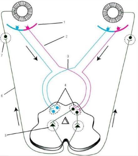
The pupillary reflex, the scheme of which is quite typical, occurs in the following order. The light that passes through the layers of the eye and is refracted into them, directly hits the retina. The photoreceptors that are located here, in this case, are the beginning of the reflex. In other words, the path of the pupil reflex begins here. The innervation of parasympathetic nerves affects the functioning of the sphincter of the eye, and the arc of the pupillary reflex contains it in its composition. The process itself is called the efferent shoulder. The so-called center of the pupillary reflex is immediately located, after which various nerves change their direction: some of them go through the legs of the brain and enter the orbit through the upper gap, while others go to the pupil sphincter. On this the path ends. That is, the pupil reflex closes. The absence of such a reaction may indicate any violations in the human body, which is why this is so important.
Pupillary reflex and signs of its defeat
When examining this reflex, several characteristics of the reaction itself are taken into account:
- the size of the narrowing of the pupil;
- the form;
- uniformity of reaction;
- pupil mobility.
There are several of the most popular pathologies, indicating that the pupil and accommodation reflexes are impaired, which indicates a malfunction in the body:
- Amavrotic stillness of the pupils. This phenomenon is a direct reaction in the illumination of the blind eye and a friendly reaction if vision problems are not observed. The reasons most often are a variety of diseases of the retina and the visual pathway. If the immobility is one-sided, is a consequence of amaurosis (retinal damage) and is combined with dilated pupil, although insignificant, then there is a possibility of anisocoria (pupils become different sizes). With such a violation, other pupillary reactions are not affected in any way. If amaurosis develops on two sides (that is, both eyes are affected at the same time), then the pupils do not react in any way and even when exposed to sunlight remain dilated, that is, the pupil reflex is completely absent.
- Another type of amavrotic immobility of the pupils is the hemianopic immobility of the pupil. Perhaps there is a lesion of the optic tract itself, which is accompanied by hemianopsia, that is, blindness of half of the visual field, which is expressed by the absence of a pupil reflex in both eyes.
- Reflex immobility or Robertson syndrome. It consists in the complete absence of both direct and friendly reaction of the pupils. However, unlike the previous type of lesion, the reaction to convergence (narrowing of the pupils if the gaze is focused on a certain point) and accommodation (changing the external conditions in which the person is) is not broken. This symptom is due to the fact that there are changes in the parasympathetic innervation of the eye in the case when there are lesions of the parasympathetic nucleus, its fibers. This syndrome may indicate the presence of a severe stage of syphilis of the nervous system, less often the syndrome reports encephalitis, a brain tumor (namely in the legs), as well as a traumatic brain injury.
- Absolute, or complete immobility of the pupil (that is, it does not narrow, and does not expand at all). When exposed to a pupil by a beam of light rays, the absence of both a direct and friendly reaction to the stimulus is diagnosed. A similar reaction develops not instantly, but gradually. As a rule, it begins with a violation of physiological pupil reactions - mydriasis (dilated pupil), lack of pupil mobility.
The causes may be inflammatory processes in the nucleus, root or trunk of the nerve responsible for eye movements, the focus in the ciliary body, tumors, abscesses of the posterior ciliary nerves.