From childhood we are told not to joke with sight. Indeed, the eye is a very sensitive mechanism, which is easy to damage. One of the serious diseases associated with vision is retinal detachment. What it is, how to treat it and what it can lead to is described below.
What is the retina?
Before talking about retinal detachment, you need to understand what the retina is. Let us recall the course of the world around us in elementary school: the retina is such a part of our eye that the lens conveys an image. The retina perceives what it sees, converts it into nerve impulses, sends them to the brain - and we understand that we saw a cow, an apple or a television. In other words, the retina is a separate layer of the eye, very thin, which is the first to receive information about the visual perception of an object. She acts as a kind of "courier", a transmitter of information - receives it from the outside and sends it further to the brain.
The retina has a very complex structure - it has as many as ten separate layers, the most important of which, perhaps, are the first two - the pigment epithelium (responsible for the entry of certain substances into the retina from the capillaries) and photoreceptors, or, in other words, rods and cones. With the help of the first we are able to see in the dark, they are responsible for black and white. The second ones help to see the whole spectrum of multi-colored paints; they are active in bright light.
Retinal detachment: what is it?
So, the retina receives and transmits information about what we see. All ten layers of the retina (including rods and cones) take an active part in this . But it happens that these photoreceptors are separated from the layer of pigment epithelium. This happens if fluid builds up between these layers. In this case, it falls into other layers of the retina. Because of this, the outer layers of the retina cease to receive food, the eye loses sight. Thus, retinal detachment is a serious disease that, if measures are not taken in time, can lead to blindness.
Only at the beginning of the XVIII century, the term "retinal detachment" began to be used in medicine, however, such a diagnosis was impossible to establish due to the lack of necessary devices for another whole century and a half. Now it is known that those who suffer from myopia, diabetes mellitus or vascular diseases, as well as those who have suffered eye injuries, are at greater risk of this disease. However, it must be remembered that traumatic tearing of the retina occurs in approximately 6% of the world's population and only in isolated cases leads to delamination.
Types of retinal detachment
There are 5 types of retinal detachment: traumatic, traction, exudative, primary or secondary. Primary detachment occurs due to rupture of the retina, secondary - due to all kinds of inflammatory processes in the eye, including tumors. And traumatic is, as the name suggests, the result of an eye injury. Exudative detachment is called when there is no retinal rupture, but liquid has accumulated under it. And finally, tractional detachment is one in which retinal tension occurs.
It is imperative for a specialist to know what kind of retinal detachment happened, as this will help determine the further course of treatment.
Why does the retina exfoliate?
The reasons for retinal detachment are quite simple and commonplace. First of all, these are retinal breaks, which are already mentioned above. These tears appear due to inflammation of the lining of the eye, severe myopia, hemorrhages in the eyes, great physical exertion, and so on. In addition, an eye injury can become the cause of retinal detachment - even if it was a long time ago, after a while it can make itself felt. In order to detect the problem in time and avoid even greater troubles, it is necessary to regularly visit an ophthalmologist. By the way, the older the person, the greater the risk of retinal detachment. And if a similar problem arose in the patient in one eye, the likelihood of developing the disease in the other is also high.
Symptoms
How to recognize what happened? There are several sure signs. First, the symptoms of retinal detachment in the early stages include the so-called light phenomena - sparks and flashes begin to flash before the eyes. This suggests that photoreceptors are irritated. You must not miss this signal and consult a specialist in time. Still symptoms of retinal detachment are floating circles, dots, a veil in front of the eyes. This is a sign of damage to the vessels of the retina. Often, the described symptoms appear at the same time, but it happens that the outbreaks are a couple of days ahead of the circles.

What's next? Further, if you do not pay attention to the signals sent by the body and ignore them, retinal detachment will progress. Gradually, deterioration will begin. A curtain appears before my eyes - first on the sides, so lateral vision is lost, then it spreads to the whole eye. The symptoms of retinal detachment include loss of visual acuity - everything will begin to blur before the eyes, objects will lose their shape, become fuzzy, ghostly. All this leads to the worst that can happen with vision - to total blindness.
It may take several months from the first sign to the last, or maybe one week. It all depends on where the retina ruptured or the eye was injured. By the way, in the mornings, even after detachment of the retina, vision is better than in the evenings - all because in a horizontal position (if you sleep on your back), the fluid in the eye dissolves somewhat, allowing the retina to partially return to its place. However, this only happens in the first days after exfoliation - if the situation has dragged on, the retina has already lost its shape and is not able to lie on its own to the right place.
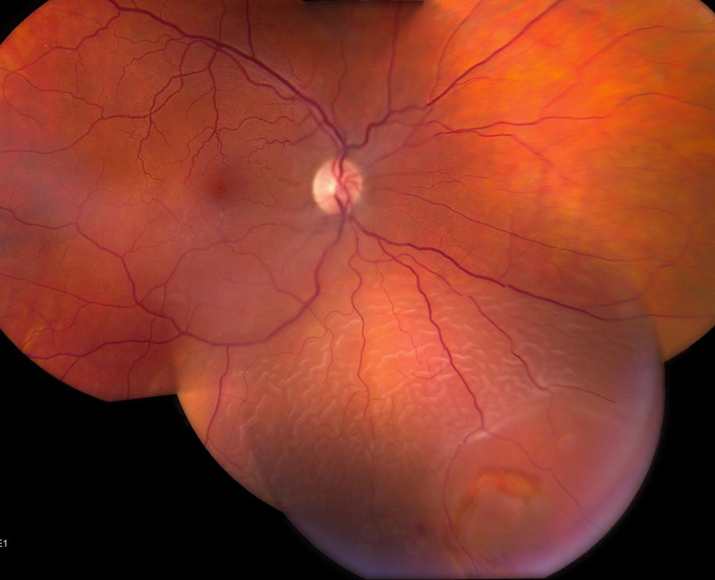
Detachment Diagnostics
Suppose a person suspected retinal detachment. What to do, what to do? Immediately run to the doctor for examination - the only way, through diagnosis, you can confirm or deny the fears. The sooner the diagnosis is made, the better - as already mentioned, timely measures will help to get by with little blood and save eyesight.
On the examination of the eye, the patient will see the visual fields to assess the condition of the retina on the periphery; examine the fundus, determine visual acuity, find out how viable retinal nerve cells are; measure intraocular pressure and so on. There are several examination methods, but ophthalmoscopy (examination of the fundus) is considered to be the main one. This diagnostic method most accurately allows you to determine whether there is a violation, and if so, to what type it belongs.
Retinal Detachment: Treatment
So, the diagnosis is clear - exfoliation. Now treatment is needed. What will it be?
There are several treatments available. The first is folk methods, the second is surgical intervention. About folk remedies we will go a little lower, but for now we should dwell on various surgical procedures. Their goal is the ability to fit the retina to the desired tissues of the eye, that is, return it to its place. Such methods include, for example, laser treatment, which strengthens the retina and limits the gap.
Treatment of retinal detachment is also possible with the help of vitrectomy - this is the removal of the vitreous from the eye and the temporary introduction of a special gas to facilitate retinal fit. Another surgical method is freezing a damaged retina, the so-called gluing of places of its rupture. This method is scientifically called cryopexy.
Using sclerotherapy, a piece of elastic plastic is placed on the outer layer of the eye so that pressure decreases on the retina and there are no new tears. And the retinopexy method allows you to introduce air into the eye, which prevents the accumulation of fluid under the damaged areas of the retina.
Laser treatment
Let us dwell on laser treatment in more detail. The laser creates adhesions between the retina and choroid, burning the retina with light. Scientifically, this method is called laser coagulation. It is carried out under anesthesia (as a rule, they give local anesthesia - they inject an anesthetic in solution). The operation is as follows: install a special three-dimensional lens on the eye, with which light rays can be projected onto absolutely any part of the fundus. The laser is sent to the necessary places, eliminating gaps, fastening the retina and choroid.
Although the operation takes a fairly small amount of time, the resulting adhesions, however, it takes about two weeks to become durable. When this happens, the retinal detachment operation is considered successful.
However, you need to be prepared for possible complications. This happens infrequently, only if the area of the treated surface was too large (and this is optional). With appropriate treatment, these complications do not cause much concern and go away in a few days.
Laser intervention is carried out not only for medical, but also for preventive purposes. This is especially true for people who are in the so-called risk group - that is, those who have an increased risk of retinal detachment. After this procedure, it is necessary to visit a specialist at least once every six months with the aim of a routine examination of the fundus. If you regularly perform these simple manipulations, the risk of developing the disease will significantly decrease.
Peeling operations
The sooner you see a doctor, the higher the likelihood of a maximum cure and a successful intervention. Experts warn that it is possible to restore vision completely and completely only if the detachment of the retina has not reached the center. In the opposite situation, vision will not be the same.
Before the operation, it will be necessary to pass tests. This is a general blood test, blood type and Rh factor, a biochemical blood test, an HIV test, a general urinalysis, cardiogram, fluorography. In addition, it is required to undergo consultations of narrow specialists: a dentist, an otolaryngologist, an endocrinologist (if there is diabetes or problems with the thyroid gland), as well as a therapist. When registered with a neurologist, dermatologist and the like, you should also visit them.

It is important to remember that retinal detachment surgery is possible if more than a year has passed since the vision deterioration. Rather, it is possible to carry out the intervention at a later date, but no one will guarantee the return of vision in such circumstances. Another important factor is that after surgery, retinal detachment often increases myopia or astigmatism. In some cases, there are relapses - exfoliation occurs again. The second operation, unfortunately, may also not be effective.
Any surgical intervention for detachment of the retina is painless, since, as already mentioned, it is performed with an anesthetic. Also, they are all safe, as the equipment for such operations is the latest. And, perhaps, the main plus - they are short, do not require a hospital stay. On average, retinal surgery lasts from forty minutes to one and a half hours.
After operation
Within a month after the intervention, it is not recommended to go to the bathhouse, sauna or pool. Depending on how the operation was and how complicated, physical activity is also limited - for a minimum of a month, a maximum of a year. In addition, at least a day immediately after the operation, a mandatory bed rest is prescribed (it, by the way, must be observed before the procedure).
The attending physician will prescribe the necessary medications, which must be taken without omissions. Also, you will not be able to lean forward, you will need to constantly monitor the position of the head, wear sunglasses. It is advisable to take care not to undergo colds.
It is not necessary to think that, having opened his eyes after the operation, a person will immediately begin to see as before, or at least better. The restoration of the functions of vision takes a certain period of time, as a rule, even several months.
Folk methods
To folk remedies include all kinds of conspiracies, compresses, juices and decoctions, herbal infusions and the like. Unfortunately, no matter how people believe in the effectiveness of these methods, in the treatment of retinal detachment, they are useless and powerless.
Eye drops, Chinese medicine, acupuncture, eye exercises and so on will also not have the desired result. Retinal detachment is a serious disease that can only be removed by surgery and nothing more.
Preventative measures
It has long been known: it is easier to prevent a disease than to treat it. And in order to prevent possible detachment of the retina, you need to visit an optometrist once at least six months. In the event that the eye was injured, further observation by a doctor is imperative.
Interesting Facts
- The human eye weighs approximately 7 grams.
- The most rare eye color is green (only 2% of the world's inhabitants have it).
- And only 1% of the world's population boasts multi-colored eyes.
- We blink every 4 seconds.
- The cornea of the human eye is terribly similar to the cornea of a shark's eye.
- A person perceives only red, yellow and blue colors, the rest are a combination of the above.
- Aphakia is a disease in which a person does not have a lens.
- If a person is afraid of the eyes, this is called ommatophobia.
- Newborn babies see at a distance of about 30-40 centimeters: it is at this distance during breastfeeding that the mother’s face is located from their eyes.
- Brown eyes are actually blue, brown pigment made them.
Our eyes serve us as a faithful service, but require careful attitude and care. Therefore, do not ignore them if you have any vision problems.