Eye biomicroscopy is a modern diagnostic method for researching vision, carried out using a special device - a slit lamp. A special lamp consists of a light source, the brightness of which can vary, and a stereoscopic microscope. Using the biomicroscopy method, an anterior segment of the eye is examined.
Indications
This method is used by an ophthalmologist in conjunction with a standard visual acuity test and fundus diagnosis. Biomicroscopy is also used if a person suspects that he has an eye pathology. Deviations in which the doctor prescribes this examination include: conjunctivitis, inflammation, foreign bodies in the eye, neoplasms, keratitis, uveitis, dystrophy, opacities, cataracts and so on. Eye biomicroscopy is prescribed during the examination of vision before and after surgical treatment of the eye. Also, the procedure is prescribed as an additional measure for diseases of the endocrine system.
How is the procedure going?
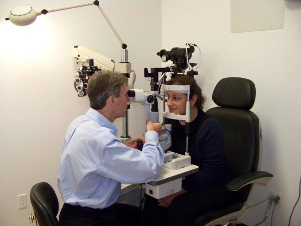
The process of biomicroscopy of the eye medium does not cause pain in the patient. A person only observes a ray of light and fulfills the doctor’s requests. The procedure does not require any special preparation and is carried out quickly. Biomicroscopy is carried out in a darkened room. The optometrist makes sure that the person takes the right position: the chin is on a special stand for the head, and the forehead is leaned against a certain place on the bar. After the patient correctly placed his head on the stand, the optometrist begins the research process. The doctor changes the direction and brightness of the light beam, while observing the reaction of the eye tissues to changes in lighting. The process of biomicroscopy of the anterior segment of the eye allows you to learn about the state of the lens and the anterior zone of the vitreous. The doctor also examines the tear film, the edges of the eyelids and eyelashes. The procedure lasts about 10 minutes. Usually this time is enough to diagnose the patient.
Ultrasound examination
The use of ultrasound as a diagnostic tool in modern ophthalmology is based on the properties of ultrasonic waves. The waves, penetrating into the soft tissues of the eye, change their shape depending on the internal structure of the eye. Based on the data on the propagation of ultrasonic waves in the eye, the optometrist can judge its structure. The eyeball consists of areas having a different structure in acoustic terms. When an ultrasonic wave hits the boundary of two sections, the process of its refraction and reflection occurs. Based on the data on wave reflection, the ophthalmologist concludes that there are pathological changes in the structure of the eyeball.
Indications for ultrasound examination
Ultrasound examination of the eye is a high-tech diagnostic method that complements the classical methods for detecting eyeball pathologies. Sonography usually follows the classic methods of examining the patient. In case of suspicion of a foreign body in the eye, the patient is first shown x-ray; and in the presence of a tumor - diaphanoscopy.
Ultrasound diagnosis of the eyeball is performed in the following cases:
- to study the angle of the anterior chamber of the eye, in particular its topography and structure;
- study of the position of the intraocular lens ;
- for carrying out measurements of retrobulbar tissues, as well as examination of the optic nerve;
- when examining the ciliary body. The membranes of the eye (vascular and reticular) are studied in situations with difficulties in the process of ophthalmoscopy;
- when determining the location of foreign bodies in the eyeball; assessment of their penetration and mobility; obtaining data on the magnetic properties of a foreign body.
Ultrasound biomicroscopy of the eye
With the advent of high-precision digital equipment, it has been possible to achieve high quality processing of echo signals obtained in the process of eye biomicroscopy. Improvements are achieved through the use of professional software. In a special program, the ophthalmologist has the opportunity to analyze the information received both during the examination and after it. The method of ultrasonic biomicroscopy owes its appearance precisely to digital technologies, since it is based on the analysis of information from the piezoelectric element of a digital probe. For the examination, sensors with a frequency of 50 MHz are used.
Methods of ultrasound examination
In ultrasound, contact and immersion methods are used.
The contact method is simpler. With this method, the probe plate is in contact with the surface of the eye. The patient is instilled with anesthetic on the eyeball, and then placed in a chair. With one hand, the ophthalmologist controls the probe, conducting the study, and the second sets up the device. The role of the contact medium with this type of examination is tear fluid.
The immersion method of eye biomicroscopy involves placing a layer of special fluid between the surface of the probe and the cornea. A special nozzle is installed on the patient’s eye, in which the probe sensor moves. Anesthesia with the immersion method is not used.